Great Lakes Bioinformatics Conference 2015
PROMOTION
GLBIO 2015 Save the Date Flyer (.pdf)
*************************************************
Image Credit: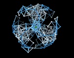
Forbes Burkowski
University of Waterloo, Canada
Insulin's Hydrophilic center
Usually one thinks of a protein as having a central hydrophobic core. In the case of insulin (PDB ID 1ZNJ) the structure is a “trimer of dimers”. Each of the trimers has a hydrophobic core but the entire structure surrounds a center that contains a hydrophilic “cage” structure containing two zinc atoms (one of which is seen in the image as a green sphere).
The image was created using VRML facilities in UCSF Chimera. Each of the residues is represented by a sphere that is given a color corresponding to the residue’s position in the Kyte-Doolittle hydrophobicity - hydrophilicity range. The corresponding color range is a linear interpolation going from orange to white to blue. Spheres having centers that are within a distance of 9 Angstroms are joined using a spindle.
For this image, all the spheres corresponding to hydrophobic residues were removed along with any incident spindles. The result reveals a compelling symmetric structure that arises from the original 3-fold symmetry of the protein.
Part of the ISMB ECCB 2013 Art and Science Exhibit
www.iscb.org/cms_addon/conferences/ismbeccb2013/artscience.php
