Art and Science Exhibition
ISMB/ECCB 2011 brings together scientists from a wide range of disciplines, including biology, medicine, computer science, mathematics and statistics. In these fields people are constantly dealing with information in visual form: from microscope images and photographs of gels to scatter plots, network graphs and phylogenetic trees, structural formulae and protein models to flow diagrams; visual aids for problem-solving are omnipresent. Some of the works of the first such exhibition at the ISMB 2008 in Toronto combine outstanding beauty and aesthetics with deep insight that perfectly proves the validity of our approach or goes beyond the problem's solution. Others were surprising and inspiring through the transition from science to art, opening our eyes and minds to reflect on the work that we are undertaking.
The Art & Science Exhibition 2011 presents the artworks that have been generated as part of research projects. They are also soliciting images resulting from creative efforts that involve scientific concepts or employ scientific tools and methods.
Art & Science Images |
Theodore Alexandrov
Univerisity of Bremen, Germany
Dennis Trede, University of Bremen, Germany
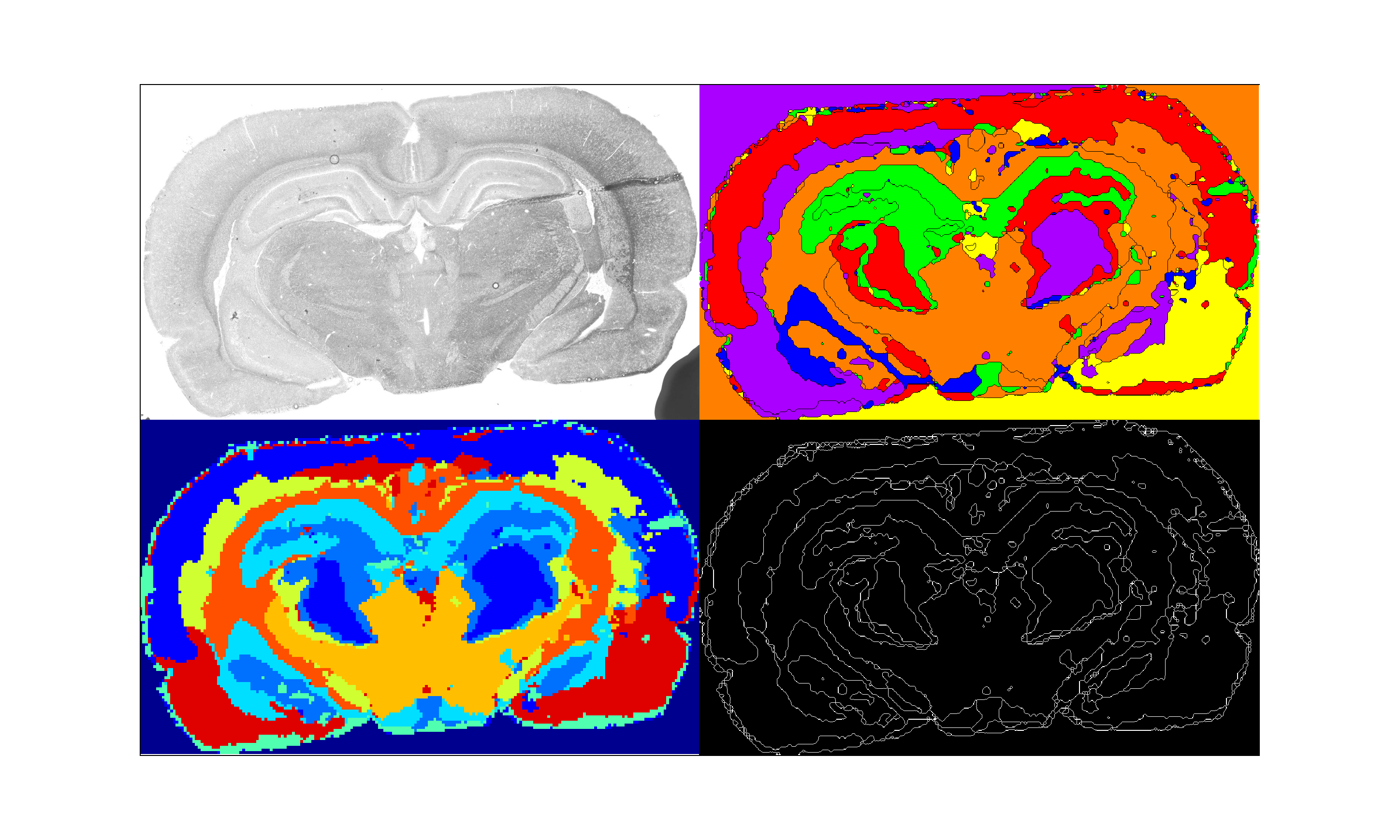
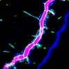
Flower-powering the Brain
This image shows an intermediate result of processing a coronal section of the rat brain measured with matrix-assisted laser desorption/ionization (MALDI) mass spectrometry imaging. The dataset has been measured at Bruker Daltonics, Bremen. The problem of interest is spatial segmentation of this dataset highlighting anatomical structure of the brain.
We were working deeply into the darkness of sophisticated mathematical image processing methods, when this lightsome image came out our view, cheerful and jazzy.
|

|
Geneviève Anhoury
France
Astronomy Gastronomy
My idea is a galaxy is a giant asleep inside an egg. When he wakes up, he separates the yolk and the white. With the white he whips up the sky, and with the yolk he fashions the earth. His hair and moustache spin the stars. With his breath, he blows the winds and the clouds. And with his voice, he makes the thunder.
The work was presented on "The 1st International Conference Art, Science and Technology: Interaction between Three Cultures", ORT Braude College, Karmiel, Israel: http://conferences.braude.ac.il/ArtIndustrial/committees.aspx
| |
Anne Bet
University of Alabam at Birmingham, United States
rEvolution
Modeled after the iconic pop art work of Andy Warhol, the duplicated image of HIV-1 virions in 293T cells was taken using transmission electron microscopy at 21000x magnification. Our lab focuses on HIV-1 peptides derived from alternative reading frames and recently showed that these “cryptic epitopes” are frequently recognized during HIV-1 infection. Identifying cryptic epitopes relies on pattern recognition techniques and prediction software, which was developed by our collaborators at Microsoft. The art is organized to highlight basic patterns and geometric shapes such as diamonds, circles, and squares. The title of the work plays upon evolution and action; despite HIV’s rapidly evolving genome and fast escape from immune pressures, collaborative research efforts forge an unstoppable revolution against an unrelenting foe. | 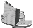 |
Anne Bet
University of Alabama at Birmingham, United States
Keys to Cryptic
Cryptic epitopes (CE) are short amino acid (aa) sequences translated from alternative reading frames that overlap, but do not contain, a known gene sequence. Recently, our lab showed that CE from both forward and reverse frames of the HIV-1 genome are recognized by T-lymphocytes in both acutely and chronically infected patients from Zambia and the US. Importantly, recognition of CE suggests HIV-1 may encode previously unknown siRNA or proteins. Understanding features of CE and their sources of generation could provide key insights to unlocking new targets for vaccine designs.
A median coding sequence (MDS; Acc: 04ZASK175B1) identified from 2, 174 available clade C sequences in the Los Alamos National Labs database was selected for analysis of open reading frames (ORFs; depicted as directional arrows based on reading sense). ORFs >25aa in length that potentially encode CE are depicted in black for HIV-1 genes and steel for predicted frames. Gated by the ORFs, the African continent is displayed as a heatmap of HIV prevalence in adults
(grey, no data; amber, 0-1.9%; ochre, 2-4.9%; orange, 5-9.9%; pink, 10-14.9%; red, >15%; UNAIDS 2009). | |
Andrzej Brodzik
MITRE, United States
Eigen-faces of Goroka
The Island of Papua is divided between the Indonesian province of West Papua (formerly Iryan Jaya) and the independent state of Papua New Guinea. Papua is the second largest island in the world. It is also one of the most remote and one of the most culturally, anthropologically and linguistically diverse places on Earth, with over 800 indigenous languages being spoken. Some of this diversity can be observed at many sing-sings, or tribal gatherings. The images included here are from the Goroka Show held in September 2009 in the village of Goroka. | |
Carlo Cannistraci
King Abdullah University for Science and Technology (KAUST), Saudi Arabia
Carlo Vittorio Cannistraci, King Abdullah University for Science and Technology (KAUST), Saudi Arabia
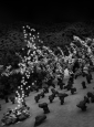
Hippocubism: Petals falling from a Blooming Tree
I love trees: from the spanning tree to the pyramidal neuron, their shapes share the same archetypal symmetry among roots and branches. And the petals are a silent interference: a patter of colors and wind.
Hippocubism is a current in the recently proposed movement: Cubism in Computational Biology [1]. Hippocampus: a virtual space where the Nature, our Great Mother, presents some of her masterpieces. My mind is in continuous search for biological patterns, and the elegance of pyramidal neurons embedded in the hippocampus was irresistible.
Here, I started from the draw of a pyramidal CA3 neuron and following the same methods presented in [1] I algorithmically forged this computational artwork.
1. Carlo Vittorio Cannistraci - A Manifesto for a Cubism movement in Computational Biology, aka CBC¿ - ISMB-ECCB 2009, Stockholm, http://www.iscb.org/ismbeccb2009/artscience.php | 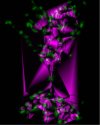 |
Anna Dehof
Saarland University, Germany
Reflections on Protein Structures
Ray tracing techniques allow unique insight into molecular structures - but they also allow to combine science and art. The image shows a human A2A adenosine receptor - a member of the famous class of G-protein coupled receptors. It was created using BALLView integrated with the real time ray tracing library RTfact. If you look closely, you can see the reflections inside the reflections inside the reflections...
The following people were directly or indirectly involved in creating this image:
Anna Katharina Dehof
Daniel Stöckel
Stefan Nickels
Sabine Müller
Iliyan Georgiev
Lukas Marsalek
Philipp Slusallek
Oliver Kohlbacher
Hans-Peter Lenhof
Andreas Hildebrandt | 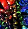 |
Jonathan Dickerson
The University of Manchester, United Kingdom
David Robertson, The University of Manchester, United Kingdom
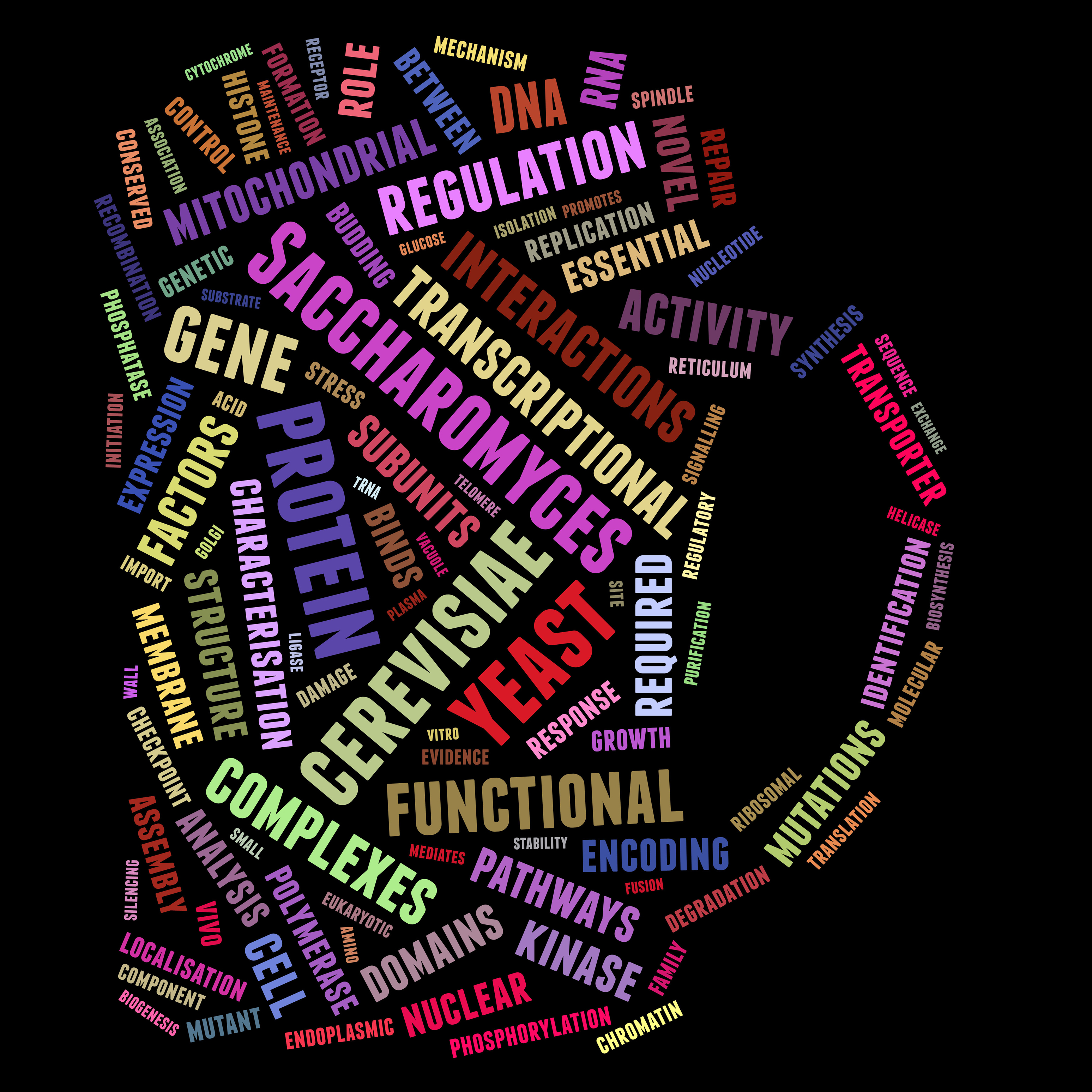
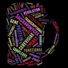
"Yeast Biology"
Populous word 'tag cloud' from 16,417 publications that chronicle 6,200 genes of the brewer's yeast Saccharomyces cerevisiae. Titles and abstracts were extracted from MEDLINE and full text, where available, was downloaded from PMC. Common and nondescript words were not considered; remaining words were stemmed, singularised and standardised (British English). Top scoring words shown; the larger the font - the greater the global word frequency. | 
|
Jonathan Dickerson
University of Manchester, United Kingdom
Visualisation of the HIV-host protein interaction network
Characterizing the relationships and interactions between human and HIV proteins is pivotal to our understanding of HIV replication and pathogenesis and provides unique information for the development of safe therapeutic interventions. Visualisation of a network of these interactions helps us explore the plethora of data. Here, coloured spheres and black diamonds correspond to 1400 human and 9 HIV-1 proteins respectively, with grey lines corresponding to one or more unique HIV-1-to-human protein interactions. Sphere diameter and colour indicate the human protein degree (larger and cooler colour indicates more interactions). Spheres are approximately clustered based upon shared interactions, whilst overlapping spheres are a result of the visualization process and do not imply any specific relationship. Data was filtered from the HIV-1, human protein interaction database at NCBI (http://tinyurl.com/hivppi). | 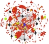
|
Yuval Eshed
Weizmann Institute of Science Rehovot, Israel
Arabidopsis carpels.
Carpels, the female organs of the plant, protect the seeds and will become the mature fruit. However, specific mutations can simulate formation of internal organs at the outer side of young carpels. In extreme cases, this can result in seeds developing inside and outside of the fruit. | |
Angelo Facchiano
Institute of Food Science, Italy
Anna Marabotti, Institute of Biomedical Technologies, Italy
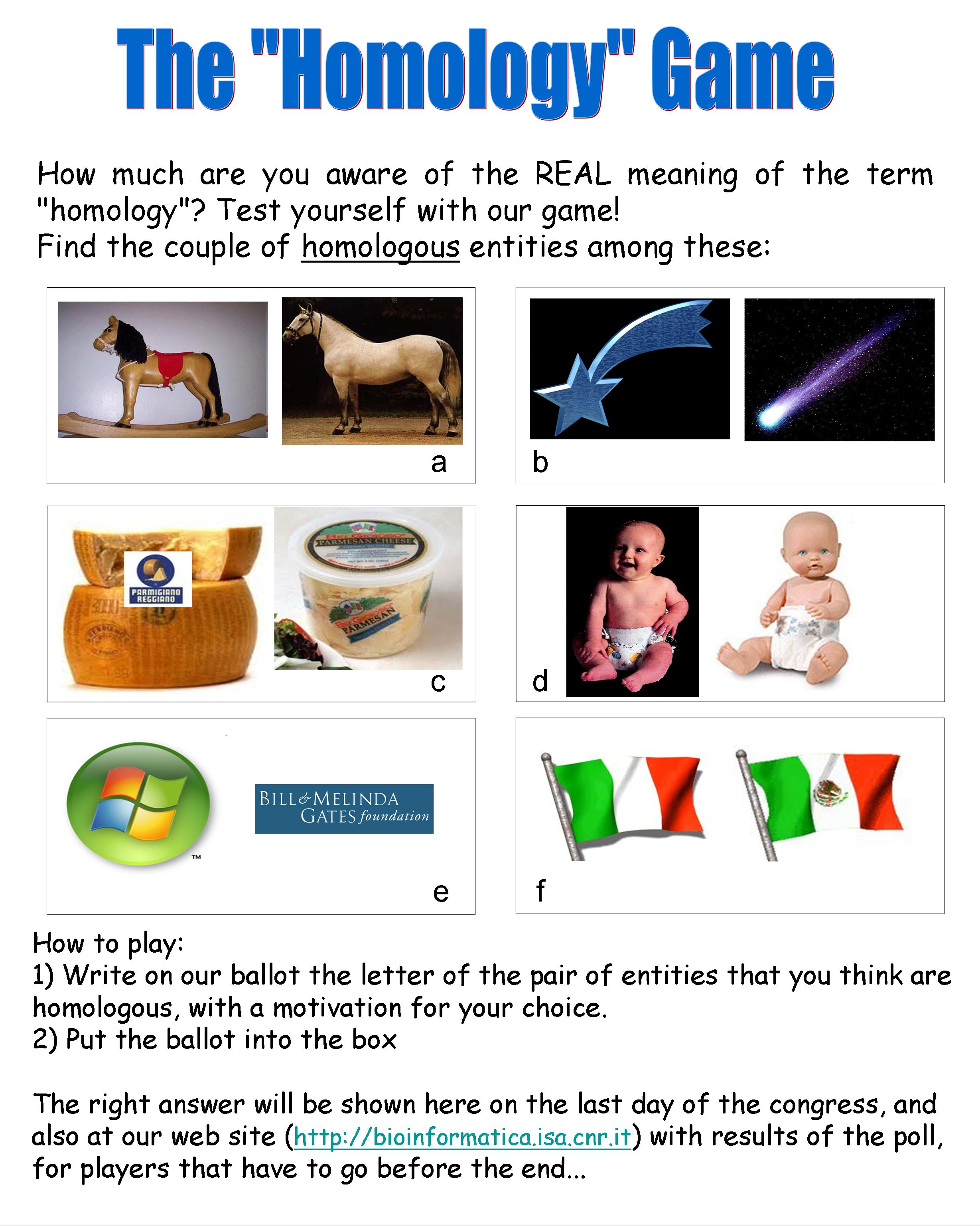
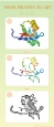
The Homology Game
It’s a long time that we are trying to aware the scientific community of the importance of the correct use of scientific terms. We are focusing our efforts on one of the most misused terms: “homology”. We present this artwork, made in form of game. The image we submit is a quiz: people should play giving us (in anonymous form) what they think is the correct answer, with a motivation for their choice. At the end of the conference, we will give the correct answer and the statistics on players’ answers. We chose a “funny way” to present this serious subject, because we think that people will focus better this problem in the future. In other words, we hope to make people “first laugh, then think” on the REAL meaning of the word “homology”. |  |
Mehrdad Garousi
Freelance fractal artist, painter, and photographer, Iran
Particulate Coliseum – The Fractal
Fractal mathematics stemming from nature behaviors and describing chaos of nature has been used as one of the very modern ways of investigation and creation in modern sciences. A substantial one is fractal art, especially newly emerged three dimensional fractal art. 3D fractals present bizarre and mostly constructible architectures that seem to belong to future. These types of massive architectures spanning different notions like chaos, order, disorder, fractional dimension, regularity, scalability, self-similarity, aesthetics, mathematics, art, science, and nature could play more applied usages in future, due to their tangible 3D structures. The work “Particulate Coliseum” discloses some of my artistic investigations to catch such an essence of future architectures among fractals.
The work was presented on "The 1st International Conference Art, Science and Technology: Interaction between Three Cultures", ORT Braude College, Karmiel, Israel: http://conferences.braude.ac.il/ArtIndustrial/committees.aspx
| |
Alex Goldshmid
Weizmann Institute of Science, Israel
Molecular domains within the Arabidopsis inflorescence meristem
Molecular markers emphasize the different functional domains in the inflorescence shoot apical meristem (SAM) of arabidopsis. YABBY gene expression (external images) marks cells which are dedicated to develop in-to organs. LATERAL SUPRESSOR gene expression (inner images) marks the boundary in between the central and the organ primordia domains, CLV3 marker expression ( the central light image) marks the central domain where population of SAM stem cells is maintained. | |
Liddy Hubbell
Jackson Laboratory, United States
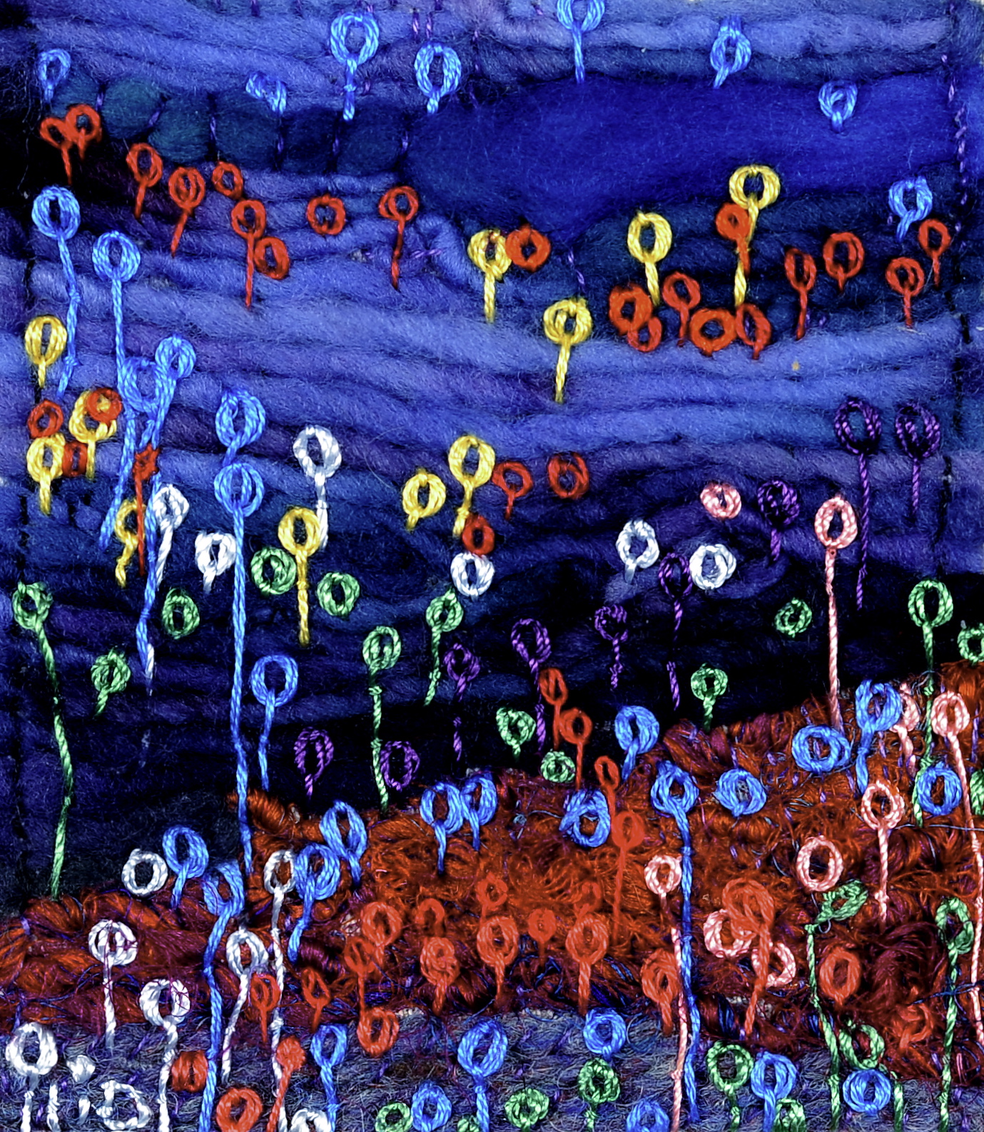
Neocortex
This collage focuses intently on the landscape of the neocortex at an unusual confluence of the visual and tactile senses. It is the complexity and richness of that juxtaposition that interests me.
This piece references work of T. Weissman, J. Lichtman and J. Sanes who used brainbow imagery to distinguish soma in a horizontally layered organization within the neocortex of a mouse. Rooted in observation, my work proceeds beyond illustration further into the abstract realm of interpretation. I use my art to convey complex scientific concepts in ways which compel fresh observation and inquiry, raise awareness of science and pose questions that we don’t normally ask.
The palm-sized work of spun silk, linen, wool and cotton might be taken as a garden, a balloon consortium, or even a landscape of tiny lollipops. Yet the fibers echo the forms of the somata and dendrites of neurons in the neocortex. The physical nature of collage materials demands attention to connections, layering and texture. The very delicacy of the threads, which crudely mimic the far finer matrices of the cells themselves, underscore the complex nature of the microscopic worlds within each of us. |  |
Maja Klevanski
University of Heidelberg, Germany
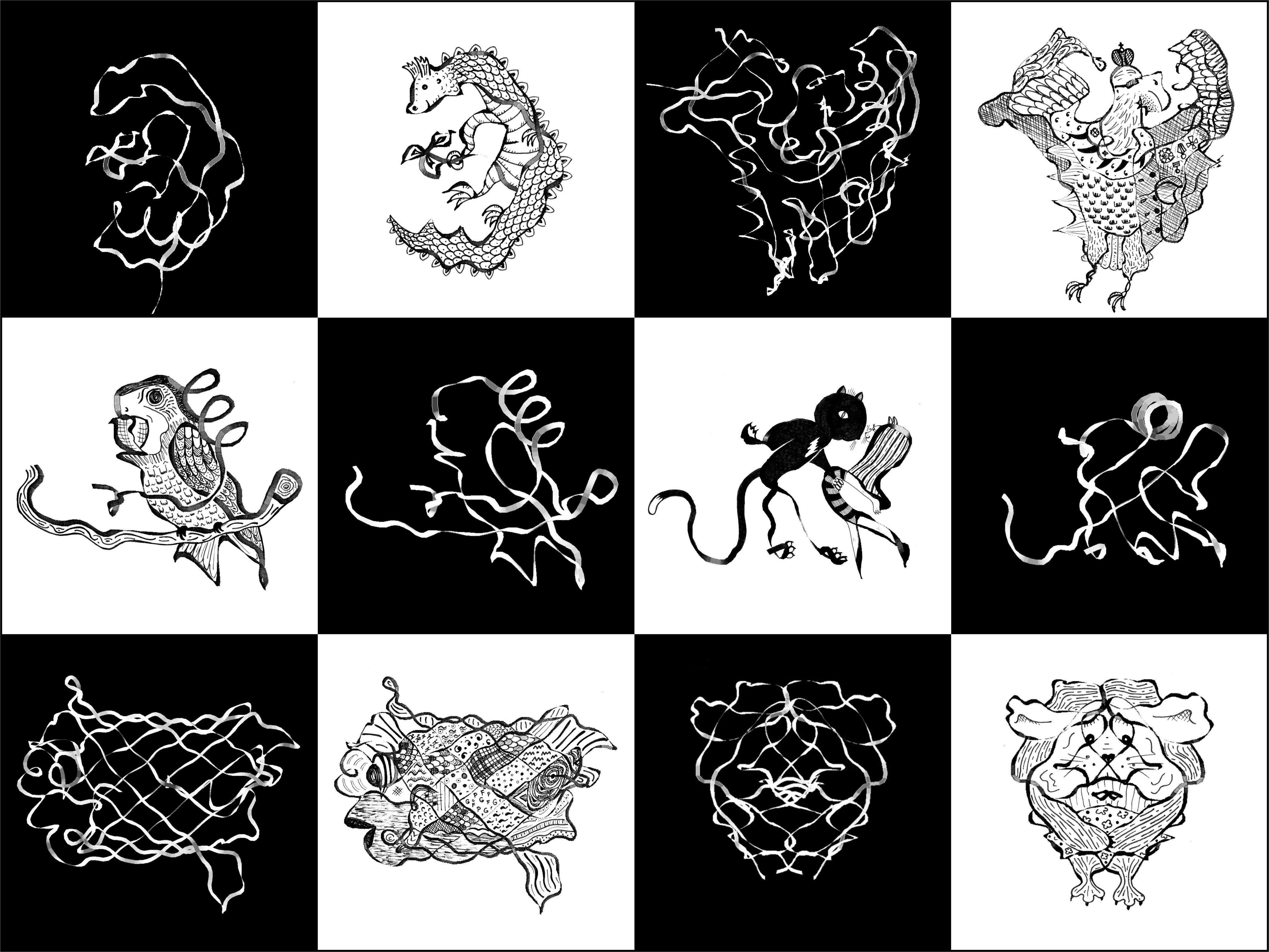
Nature Playing Chess
Explanation:
My pictures are based on contours of different proteins. I vary and rotate the three-dimensional protein image by means of a special computer software until an interesting shape appears. Subsequently, I color and design the produced snapshot so that the image I saw becomes obvious to the viewer.
Caption:
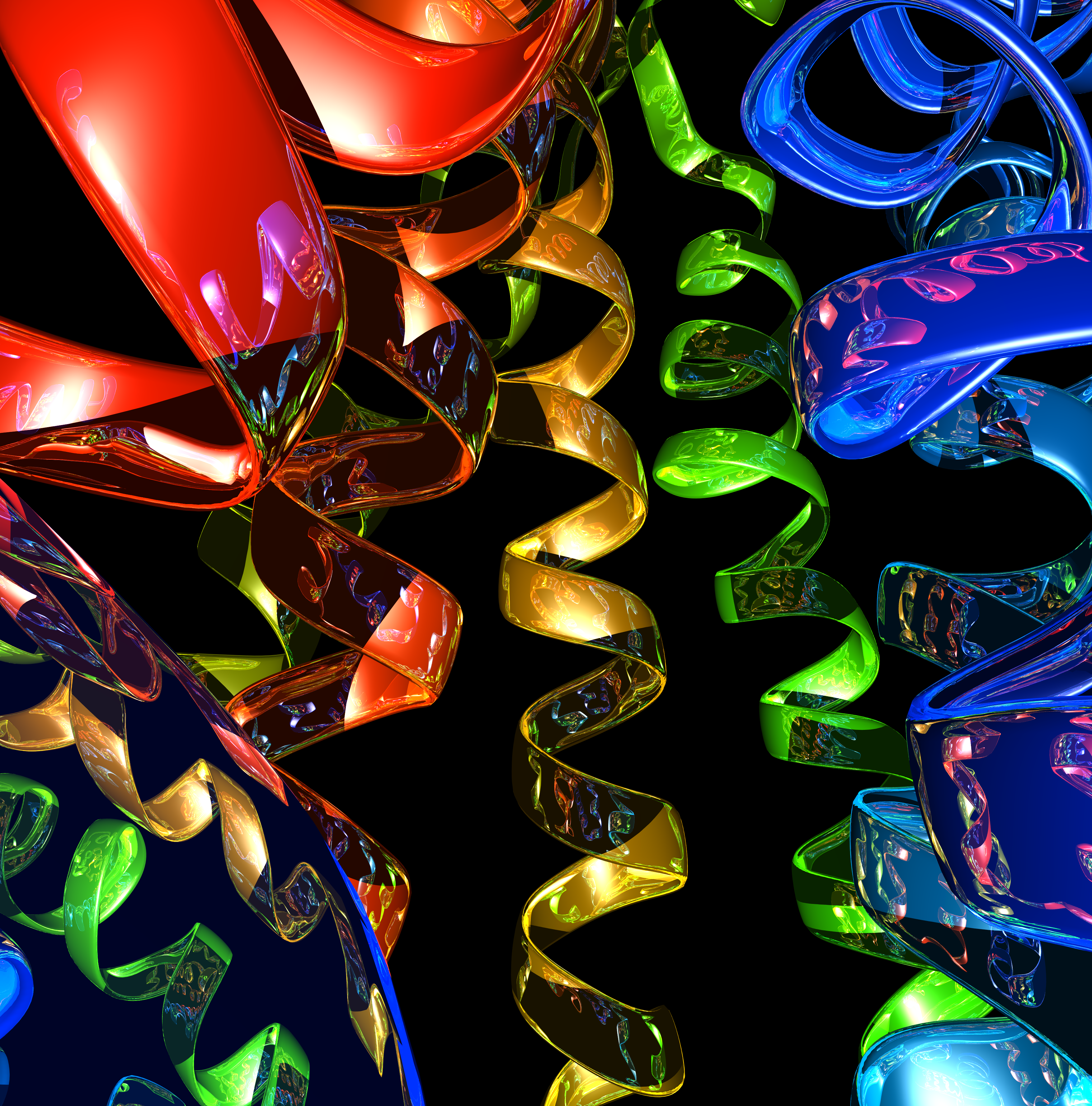
Images of twelve proteins and protein domains. The panels correspond to: N-terminal domain of APP, core domain of p53, core domain of GP120 from HIV, NC1 domain of collagen X, elastase-1, 2-nd PDZ domain of Mint1, B-chain of human insulin, PDZ domain from another perspective, GFP, alpha subunit of hemoglobin, HIV-1 protease, alpha-bungarotoxin.
Contact:
More drawings and information can be found on http://may-k.livejournal.com. | 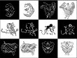 |
Maja Klevanski
University of Heidelberg, Germany
Insulin Dimer (The Fly)
My pictures are based on contours of different proteins. I vary and rotate the three-dimensional protein image by means of special computer software until an interesting shape appears. Subsequently, I color and design the produced snapshot so that the image I saw becomes obvious to the viewer.
Contact:
More drawings and information can be found on http://may-k.livejournal.com. | 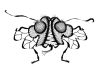
|
Maja Klevanski
University of Heidelberg, Germany
The Art Process
My pictures are based on contours of different proteins. I vary and rotate the three-dimensional protein image by means of special computer software until an interesting shape appears. Subsequently, I color and design the produced snapshot so that the image I saw becomes obvious to the viewer.
Caption:
In this example you see the 2-nd PDZ-domain of the Mint1 protein.
|
Frames PDF
|
Kaisu Koski
University of Lapland, Netherlands
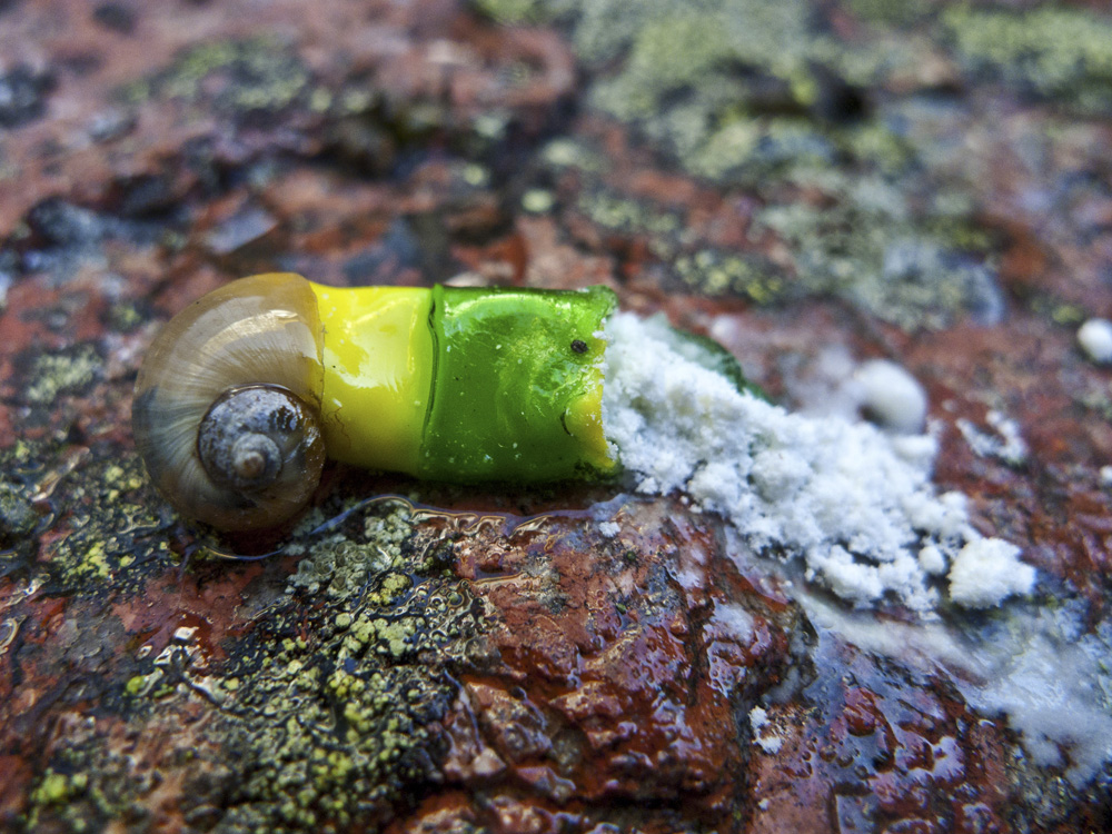
Hybrid
Hybrid merges natural and synthetic, living and dead materials in a new species. The piece envisions the future changes in organisms, resulting from the medicine residues in nature. It displays a one-shot narrative compressing a development of thousands of years. The hybrid 'snailcapsule' rests on the red-granite of the Åland Islands. |  |
Anna Lewis
University of Oxford, United Kingdom
Nick Jones, University of Oxford, United Kingdom
Mason Porter, University of Oxford, United Kingdom
Charlotte Deane, University of Oxford, United Kingdom
Multi-scale community structure of the yeast protein interaction network
Groups of proteins that are much more densely connected to each other than the rest of the protein interaction network, termed communities, are candidates for biological modules.
Here we visualize communities at multiple scales within the yeast protein interaction network. If you imagine viewing a network at arms reach, you are unable to resolve much structure (top of the image). But as you bring it closer, structure becomes apparent: you start to see a few large communities. (Closer distances are towards the bottom of the image). As you bring it yet closer, these in turn split up into smaller communities, and so on.
A horizontal slice of this image gives a network partition at a particular scale. A vertical slice gives the community membership of a particular protein at multiple scales. Blocks of color are particularly highly interconnected parts of the network. Communities below a certain size are colored black. | |
Magali Michaut
University of Toronto, Canada
Navigating the genetic landscape
Small-scale genetic interactions have been used for decades to unravel biological pathways. In recent years, experimental techniques have been developed to map them at the genome level, providing large-scale genetic interaction networks. In particular, in budding yeast the genetic network mapped by Synthetic Genetic Array (do you remember the map from last year representing The Genetic Landscape of a Cell?) was revealed as both a powerful tool to identify functional modules in the cell and a tremendous resource ready for more exploration. This image shows a view of this genetic interaction network visualized with BioLayoutExpress3D. Patterns in the genetic network can help understand the organization of biological processes (see Michaut, et al. doi:10.1371/journal.pcbi.1001092).
Image Credit: Magali Michaut, University of Toronto
CC Creative Commons Attribution License
PLoS Comput Biol 7(2): featured issue image, February 2011 | 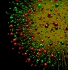 |
Vadim Mozhayskiy
UC Davis, United States
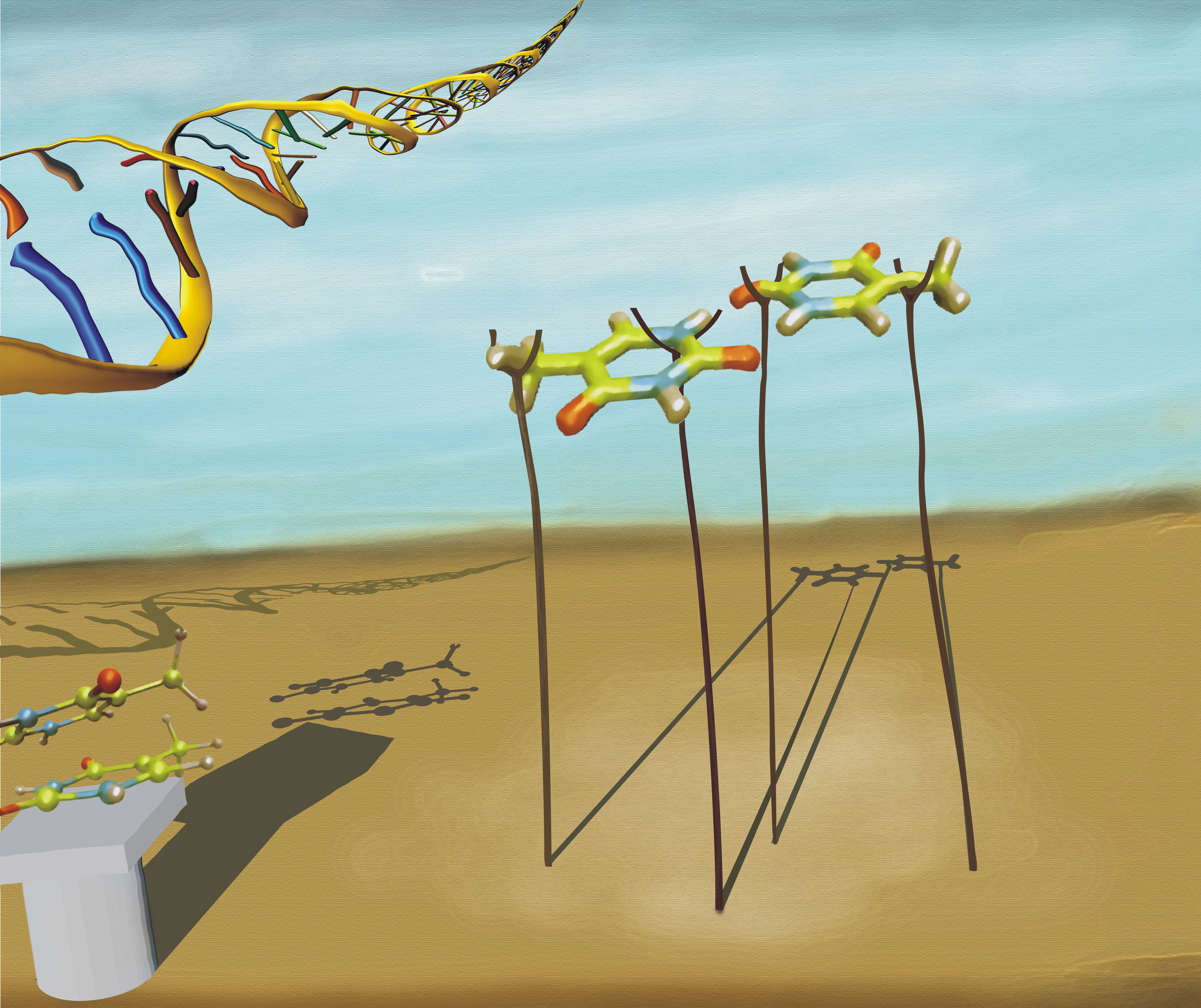
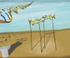
Nucleobases in a vacuum
Dimers of nucleobases in a vacuum is the simplest model system for DNA studies by high level ab initio methods and state of the art spectroscopy. However it lies miles below the Ideal -- a full-scale DNA. Therefore these studies somewhat resemble "Cargo Cults" developed by the some tribes in the 20th century after interaction with technologically advanced cultures (and this picture resembles airplanes made out of straw). However that does not belittle the quality of our state of the art research!
This work (by Vadim Mozhayskiy) was accepted as a cover art for Physical Chemistry Chemical Physics Vol. 12 Issue 10 (2010) for the paper "The effect of TT-stacking, H-bonding, and electrostatic interactions on the ionization energies of nucleic acid bases: adenine–adenine, thymine–thymine and adenine–thymine dimers" by Ksenia B. Bravaya, Oleg Kostko, Musahid Ahmed and Anna I. Krylov, Phys. Chem. Chem. Phys., 12:2292(2010) (DOI: 10.1039/b919930f).
| 
|
Noah Shamir
Israel
Frozen Vertical Flow
As a physicist, I am always fascinated by vertical ice forms. The freezing point of flowing water is determined by its hydrodynamics and specifically by the velocity and mass of the flowing water volume.
Presented is a photograph of vertical ice structures, taken in Utah, Israel, on winter 2008.
The work was presented on "The 1st International Conference Art, Science and Technology: Interaction between Three Cultures", ORT Braude College, Karmiel, Israel: http://conferences.braude.ac.il/ArtIndustrial/committees.aspx
| |
Ben-Zion Shilo
Weizmann Institute of Science, Israel
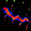
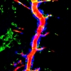
untitled
The picture shows the dorsal trunk of the Drosophila embryonic tracheal system. Apical accumulation of filamentous actin (green, observed as yellow) generated by Diaphanous/Formin is responsible for secretion to the lumen of antigens like 2A12 (red) via MyosinV-driven vesicles, but not proteins secreted through the septate junctions. Tracheal nuclei are marked by staining for Trachealess (blue). |
|
Monica Zoppè
Scientific Visualization Unit - LTGM, Italy
Lipid Raft I
The cellular membrane hosts a wide variety of proteins and different lipids. We have developed and used BioBlender, an extension of the 3D Computer Graphics package Blender, to recreate the environment of the cellular surface.
All visible objects are modeled on the basis of their atomic coordinates, with the surface imposed as a texture map, calculated with BioBledner, derived from a high resolution model: protein data are from the Protein Data Bank (K channel, CD4, Thy1/CD59, T Cell Receptor, Toll Like receptor, and others), and sugars (the little dark objects on the foreground) are built using chemical programs.
The K channel is seen releasing a few hundreds of ions, however, in the open conformation, this protein can release millions of ions per second.
|
|
Monica Zoppè
Scientific Visualization Unit - LTGM, Italy
Lipid Raft II
The cellular membrane hosts a wide variety of proteins and different lipids. We have developed and used BioBlender, an extension of the 3D Computer Graphics package Blender, to recreate the environment of the cellular surface.
All visible objects are modeled on the basis of their atomic coordinates, with the surface imposed as a texture map, calculated with BioBledner, derived from a high resolution model: protein data are from the Protein Data Bank (K channel, CD4, Thy1/CD59, T Cell Receptor, Toll Like receptor, and others), and sugars (the little dark objects on the foreground) are built using chemical programs.
The K channel is seen releasing a few hundreds of ions, however, in the open conformation, this protein can release millions of ions per second.
| |
Monica Zoppè
Scientific Visualization Unit - LTGM, Italy
Lipid Raft III
The cellular membrane hosts a wide variety of proteins and different lipids. We have developed and used BioBlender, an extension of the 3D Computer Graphics package Blender, to recreate the environment of the cellular surface.
All visible objects are modeled on the basis of their atomic coordinates, with the surface imposed as a texture map, calculated with BioBledner, derived from a high resolution model: protein data are from the Protein Data Bank (K channel, CD4, Thy1/CD59, T Cell Receptor, Toll Like receptor, and others), and sugars (the little dark objects on the foreground) are built using chemical programs.
The K channel is seen releasing a few hundreds of ions, however, in the open conformation, this protein can release millions of ions per second.
| |
Art & Science Videos |
Thomas Haschka
Université de Reims Champagne Ardenne / INSERM, France
Thrombospondin's C-Terminus
Thrombospondin's C-terminal region plays a key role in PSACH disease, as well as in cancer research. Mutations in the calcium binding stalk was shown to be responsible for PSACH a decade ago. Using molecular dynamics we are modeling the deformation of the protein in cases of calcium depletion/repletion. The movie highlights the key features of including detachment of certain parts of the lectin-like globular C-terminal as well as detachment and deformations of the wire-like stalk during calcium depletion. | 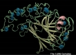
[Video]
|
Top |