


ISMB 2008 brings together scientists from a wide range of disciplines, including biology, medicine, computer science, mathematics and statistics.
In these fields we are constantly dealing with information in visual forms: from microscope images and photographs of gels to scatter plots, network graphs and phylogenetic trees,
structural formulae and protein models to flow diagrams; visual aids for problem-solving are omnipresent.
Often these visual aids are mundane and nothing more than a small clue to the solution of the problem. But then there are special ones that make the whole more than the sum of its parts. Ones that combine outstanding beauty and aesthetics with deep insight that perfectly proves the validity of the scientific approach or goes beyond the problem's solution. Ones that surprise and inspire us through the transition from science to art, ones that open our eyes and minds to reflect on the work we are doing.
The Visual Reflections on Science Exhibition@ISMB 2008 presents the artworks that have been generated as part of research projects. They are also soliciting images resulting from creative efforts that involve scientific concepts or employ scientific tools and methods.
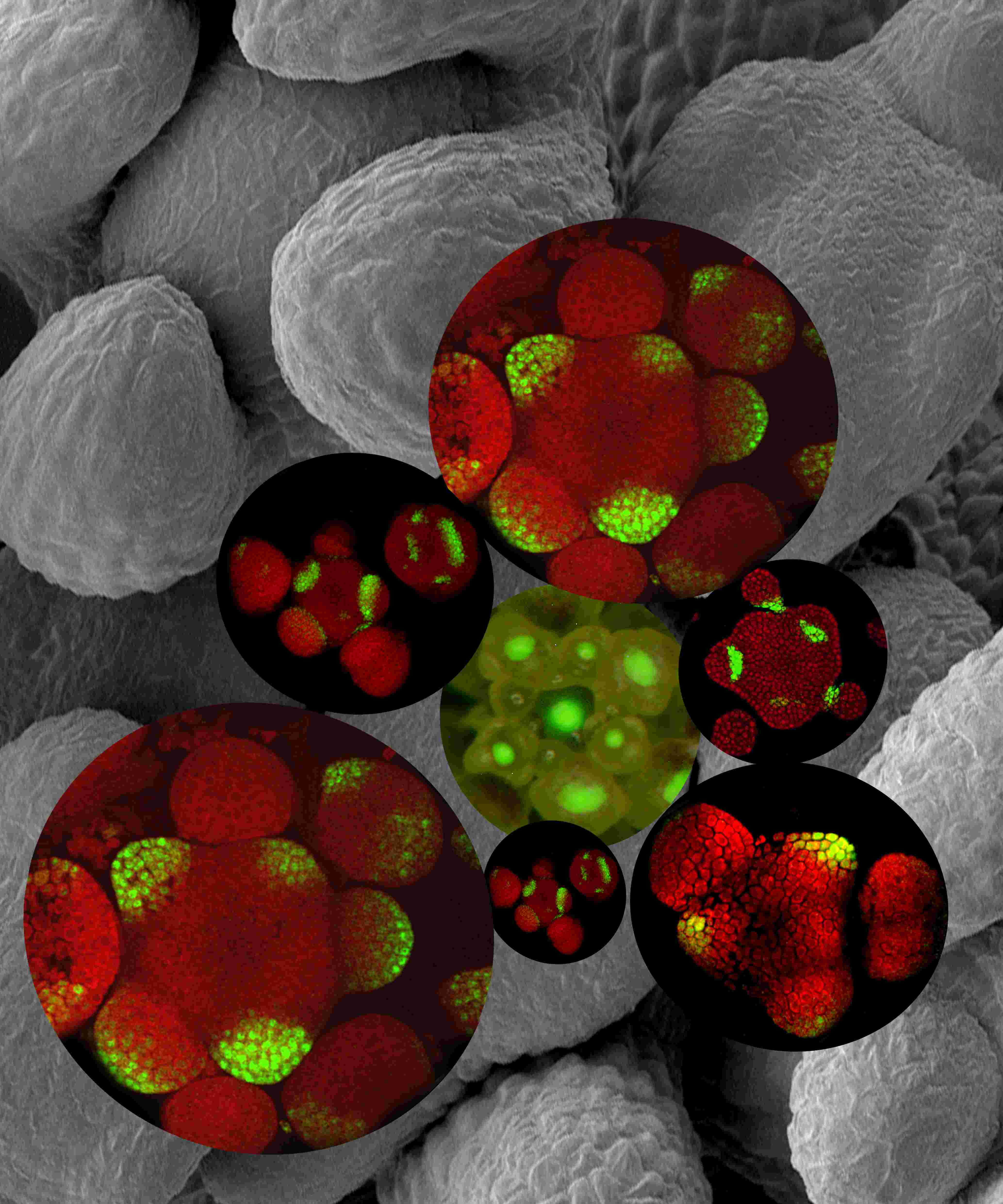
Dr.Alexander Goldshmidt Weizmann Institute of Science Rehovot, ISRAEL Molecular domains within the Arabidopsis inflorescence meristem Molecular markers emphasize the different functional domains in the inflorescence shoot apical meristem (SAM) of arabidopsis. YABBY gene expression (external images) marks cells which are dedicated to develop in-to organs. LATERAL SUPRESSOR gene expression(inner images) marks the boundary in between the central and the organ primordia domains, CLV3 marker expression ( the central light image) marks the central domain where population of SAM stem cells is maintained. |
|
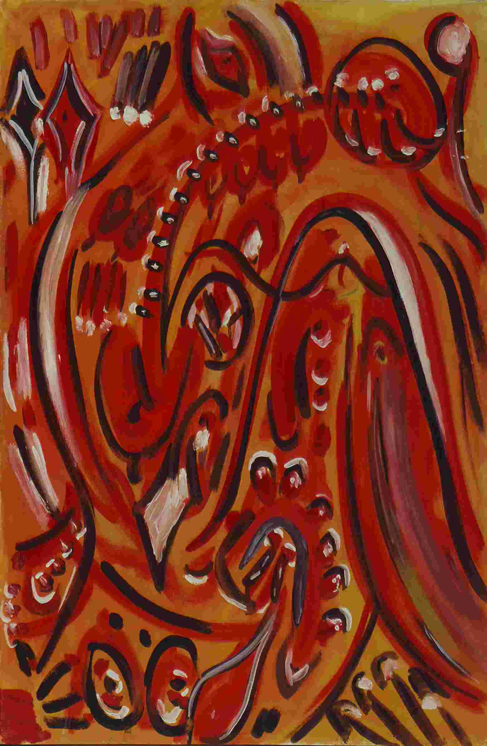
| Prof.Amos Oppenheim Hebrew University, Jerusalem, ISRAEL Bacteriophage lambda: The CII - CIII harmony [in the decision to lysogenize the host]. 1970. |
|
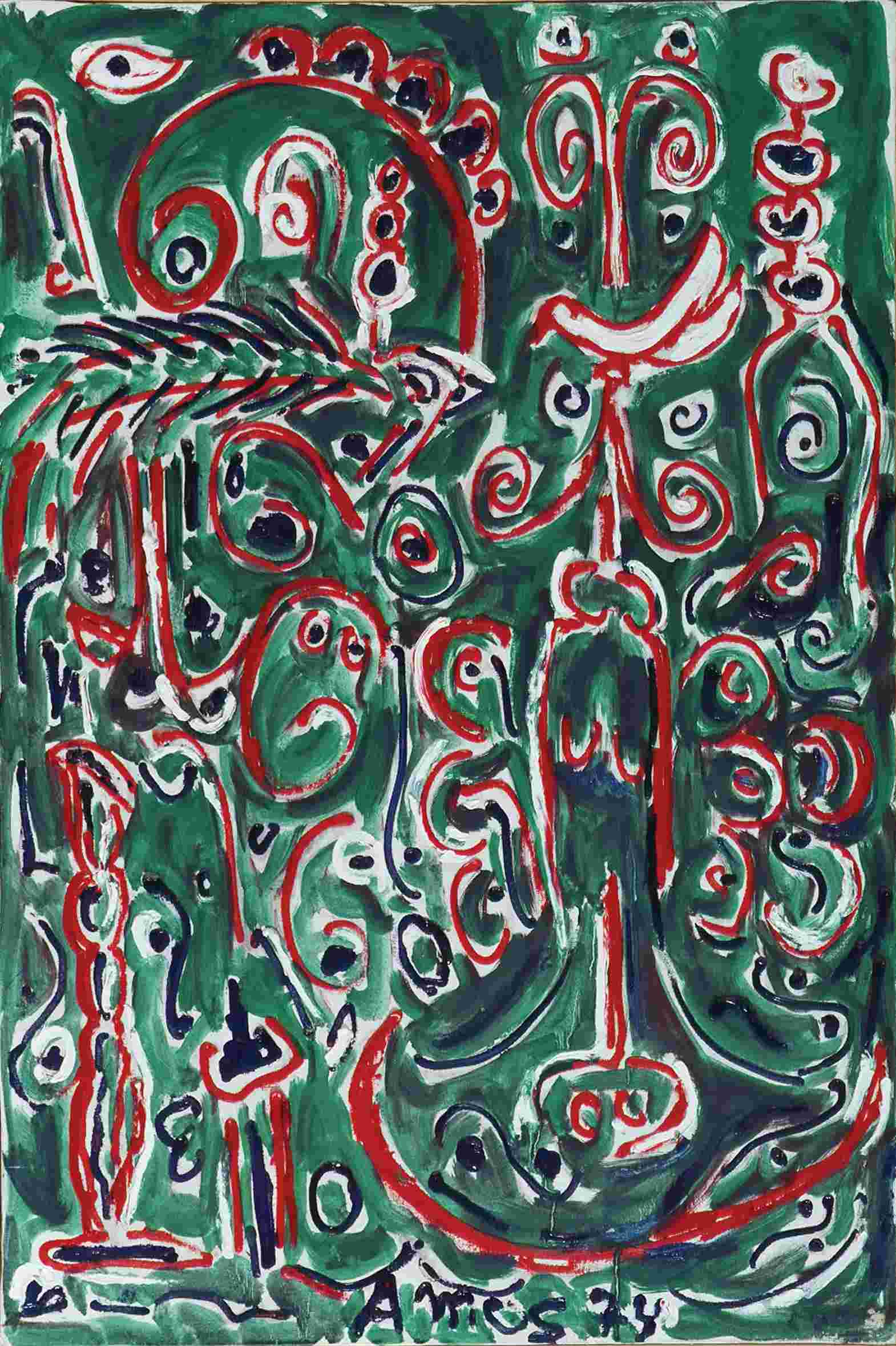
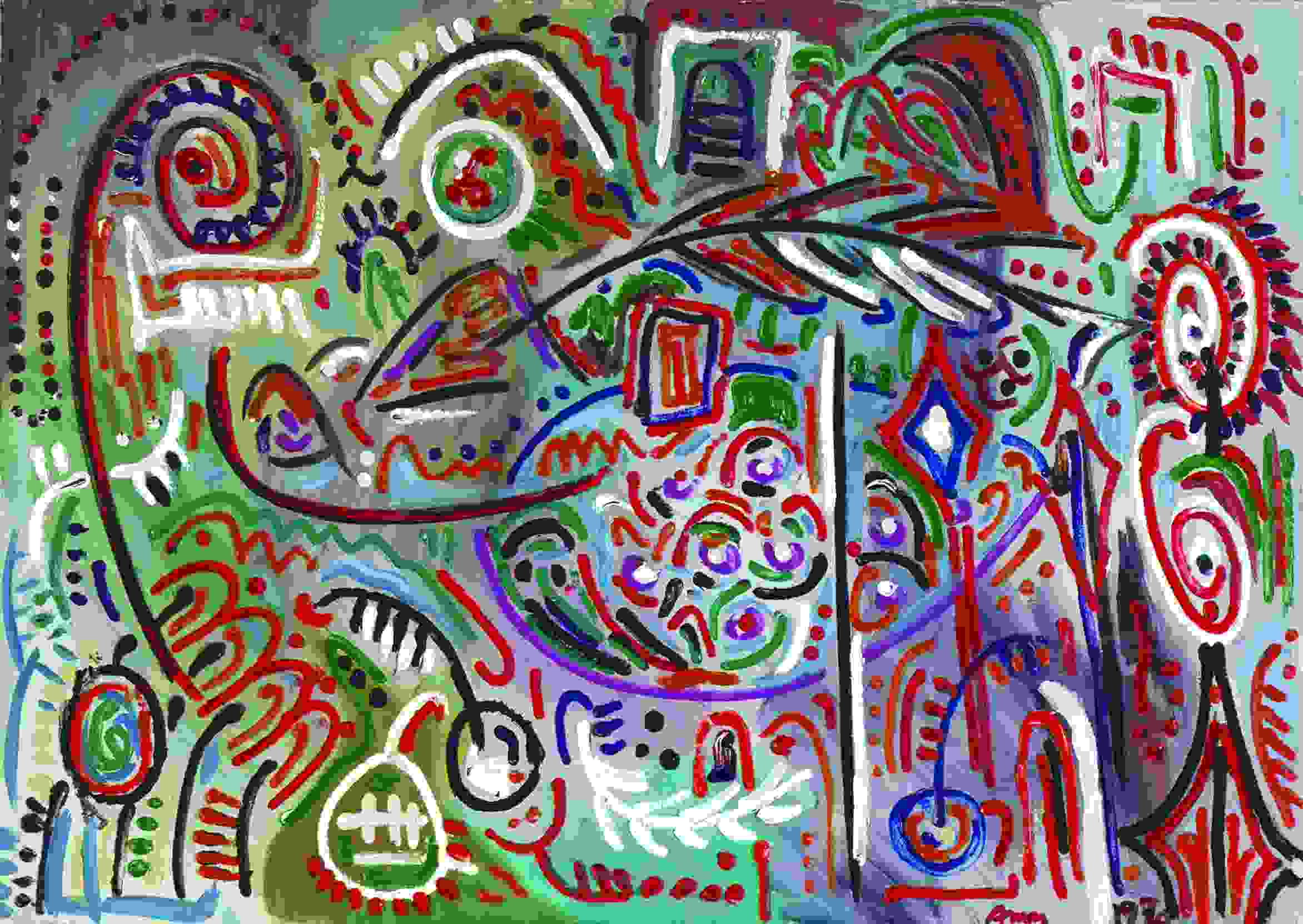
| Prof.Amos Oppenheim Hebrew University, Jerusalem, ISRAEL Evolution: from bacteria to man to peace. 1974. |
|
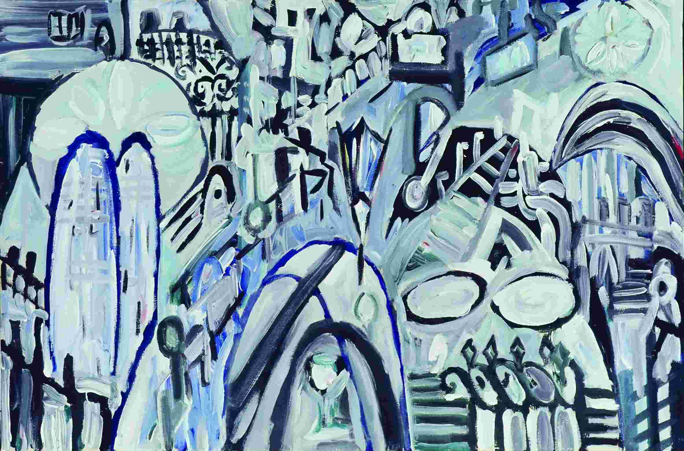
| Prof.Amos Oppenheim Hebrew University, Jerusalem, ISRAEL The gate to knowledge: Visit to Pasteur Institute. 1969. |
|
| Prof.Amos Oppenheim Hebrew University, Jerusalem, ISRAEL Gene Activation. 1970. |
|
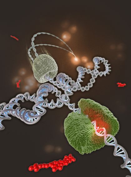
| Dr.Daniel Koster Kavli Institute of Nanoscience Delft University of Technology The Netherlands Supercoiled Topoisomerases are enzymes that act to relax supercoiling, a form of built-up strain in DNA. Topoisomerase inhibitors are important antitumour drugs, thought to act by stabilizing a covalent complex between the topoisomerase and DNA, which then sets up a road-block to the DNA replication machinery. The origin of drug efficacy in targeting topoisomerases remains poorly understood, but now a singlemolecule study of the interaction of topotecan, a drug used mainly to treat ovarian cancer and small-cell lung cancer, and a topoisomerase IB.DNA complex, has revealed more details of the process. As illustrated on the cover, positive DNA supercoils accumulate due to the action of the drug (shown red) Such overwinding of the DNA hinders an advancing DNA polymerase and may play a role in the stalling or collapse of a replication fork, ultimately leading to cell death. |
|
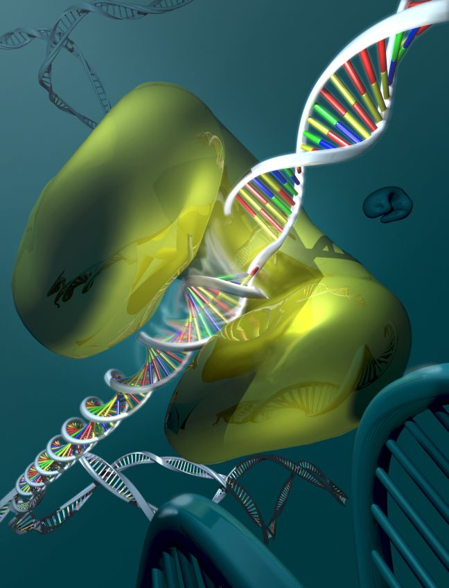
Dr.Daniel Koster Kavli Institute of Nanoscience Delft University of Technology The Netherlands Torque about DNA The manoeuvres involved in replication and transcription lead to a build-up of twisting or torsional strain in DNA, and the job of relieving the strain falls to the topoisomerases. Type IB topoisomerases (TopIBs) cut one DNA strand, allow it to swivel around its partner, then rejoin the ends. This removes both positive and negative supercoils. Direct monitoring of a single TopIB molecule now shows that, unlike other topoisomerases, it does not remove a fixed number of supercoils. Rather, the number of supercoils removed, via a mechanism involving friction between rotating DNA and the enzyme cavity, depends on the torque stored in the DNA. The introduction of friction as a factor in DNA.topoisomerase interactions may have important implications for DNA.enzyme interactions in general. The cover depicts TopIB in action. |
|
|
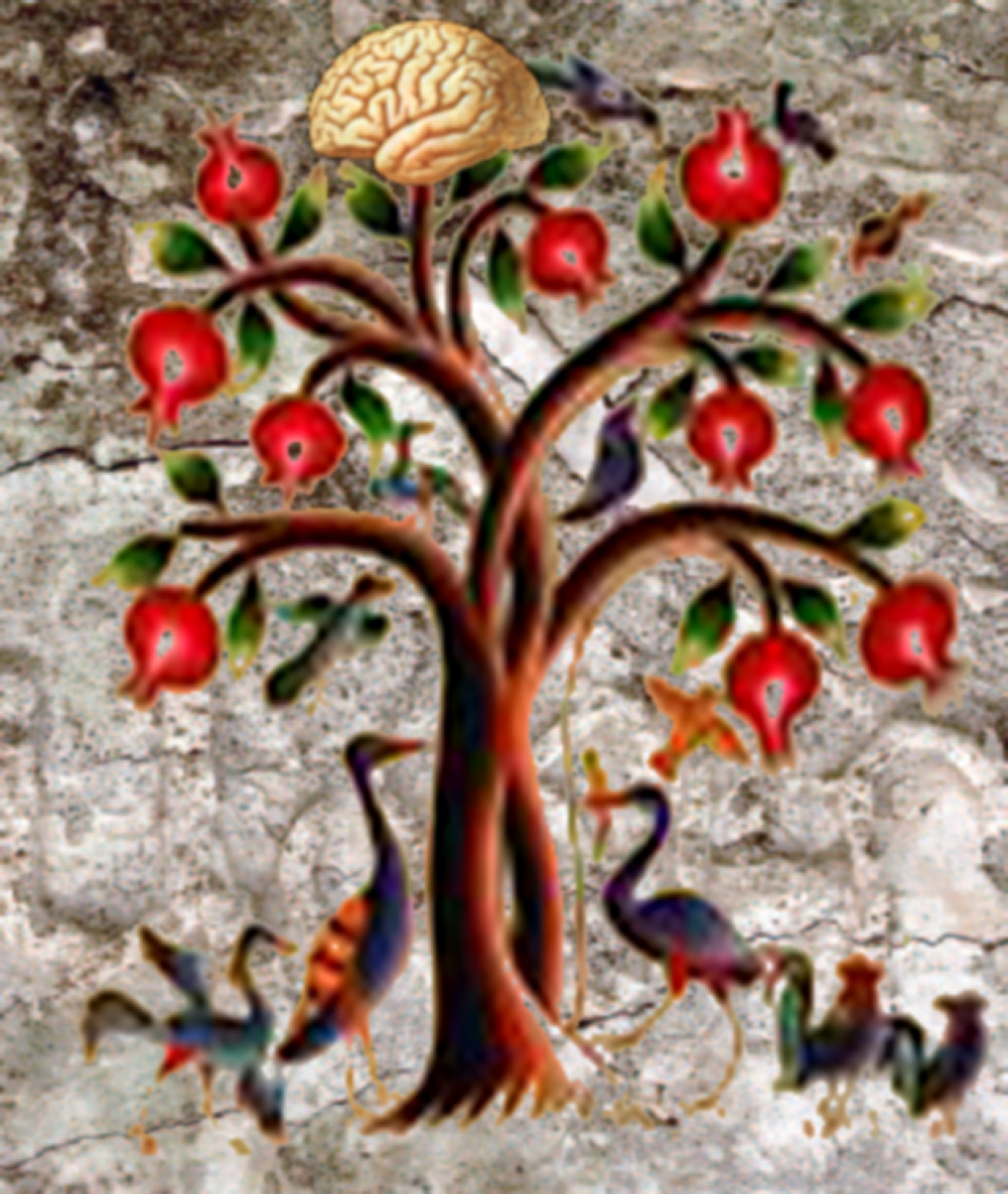
Dr. Eduard Korkotian Weizmann Institute of Science Rehovot, ISRAEL Brain through Ages The first picture is a collage made for International Organization of Armenians in Neuroscience as a symbol for this organization. It includes 3 elements. 1 - A pomegranate tree and birds, taken from a Middle Age Armenian Medical Manuscript illustrating the beauty and productivity of nature. 2 - a rock, so typical for Armenian landscape as a symbol of difficulties of scientific work and 3 - the human brain which is a product of nature on one hand and is at the top of Creation on the other hand. |
|
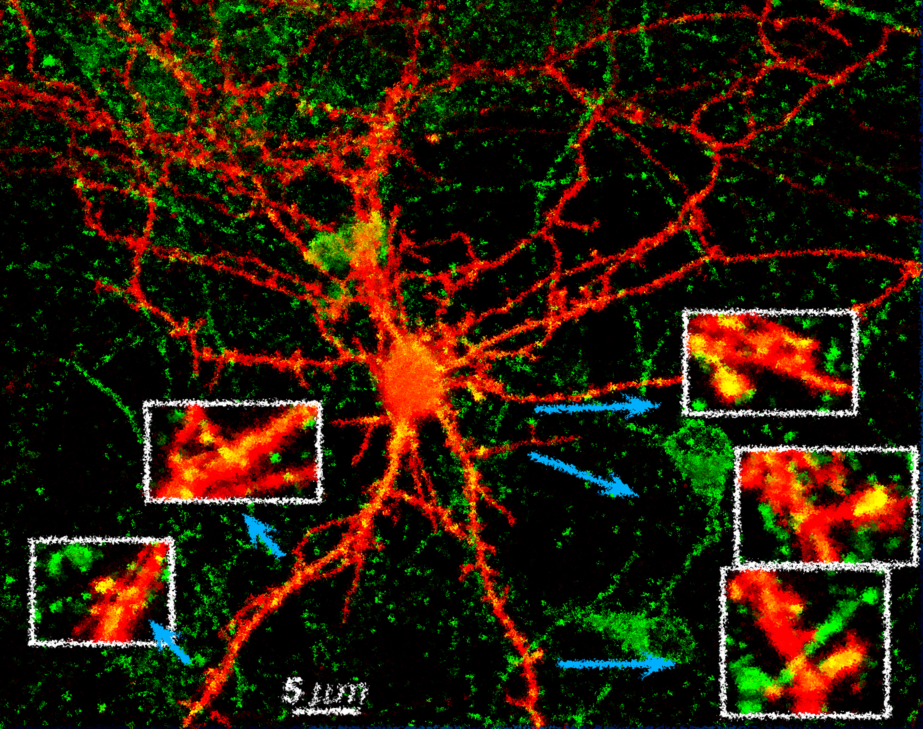
Dr.Eduard Korkotian Weizmann Institute of Science Rehovot, ISRAEL Impression of Mind The image is a hippocampal neuron, co-transfected with red fluorescent protein and green fluorescent protein-tagged AMPA receptor, responsible for neuronal activity. In white squares zoom-ins of regions labeled by arrows are shown. Original image is reconstructed from serial confocal microscope optical sections and processed with Adobe Photoshop filters. |
|
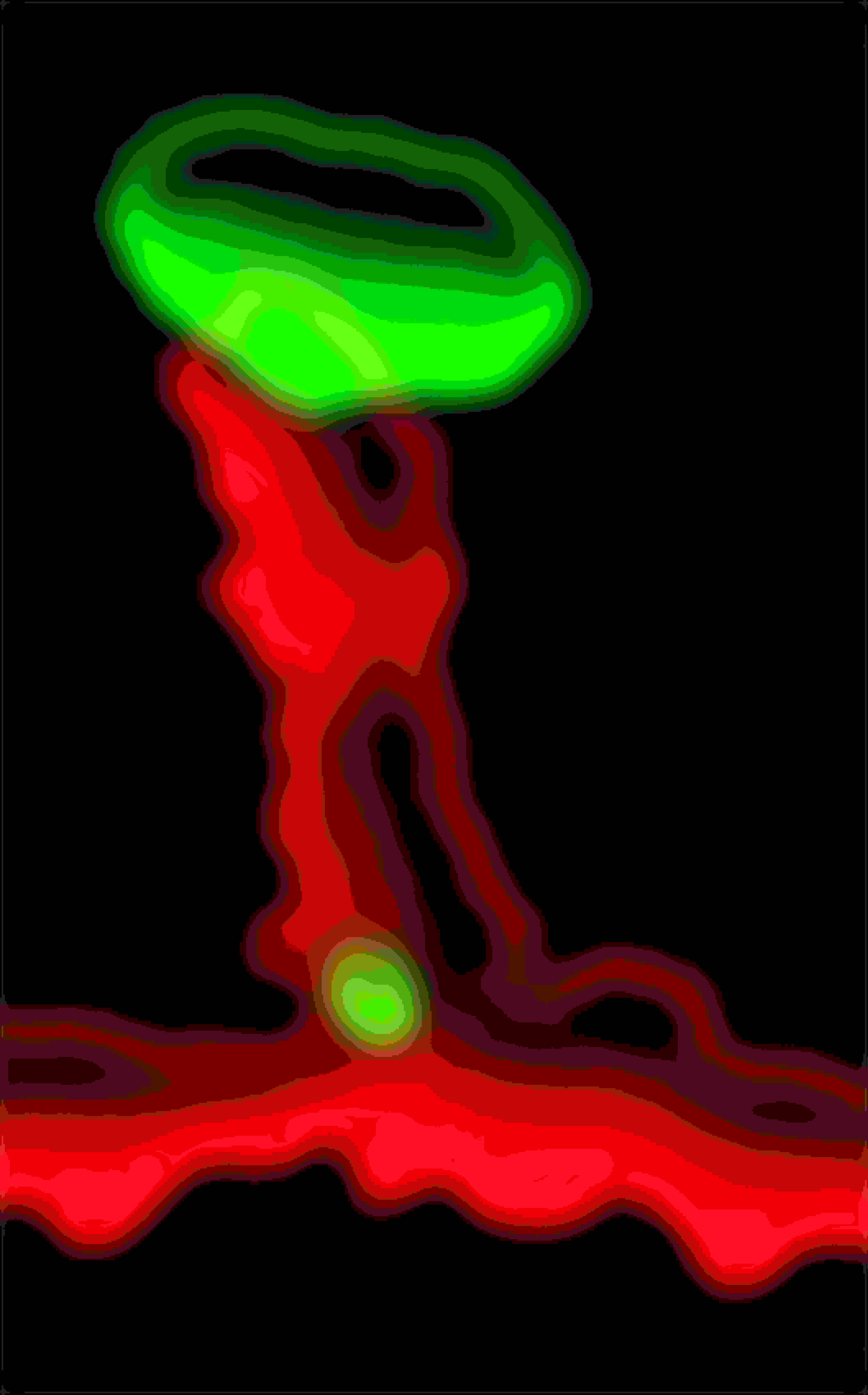
Dr.Eduard Korkotian Weizmann Institute of Science Rehovot, ISRAEL A Sinapse. Dendritic spine of the presynaptic neuron (labeled with red fluorescent protein, red) is innervated with a presynaptic terminal (labeled with green fluorescent protein tagged synapse-specific protein, green). The size of a synapse is about 1 micron. |
|
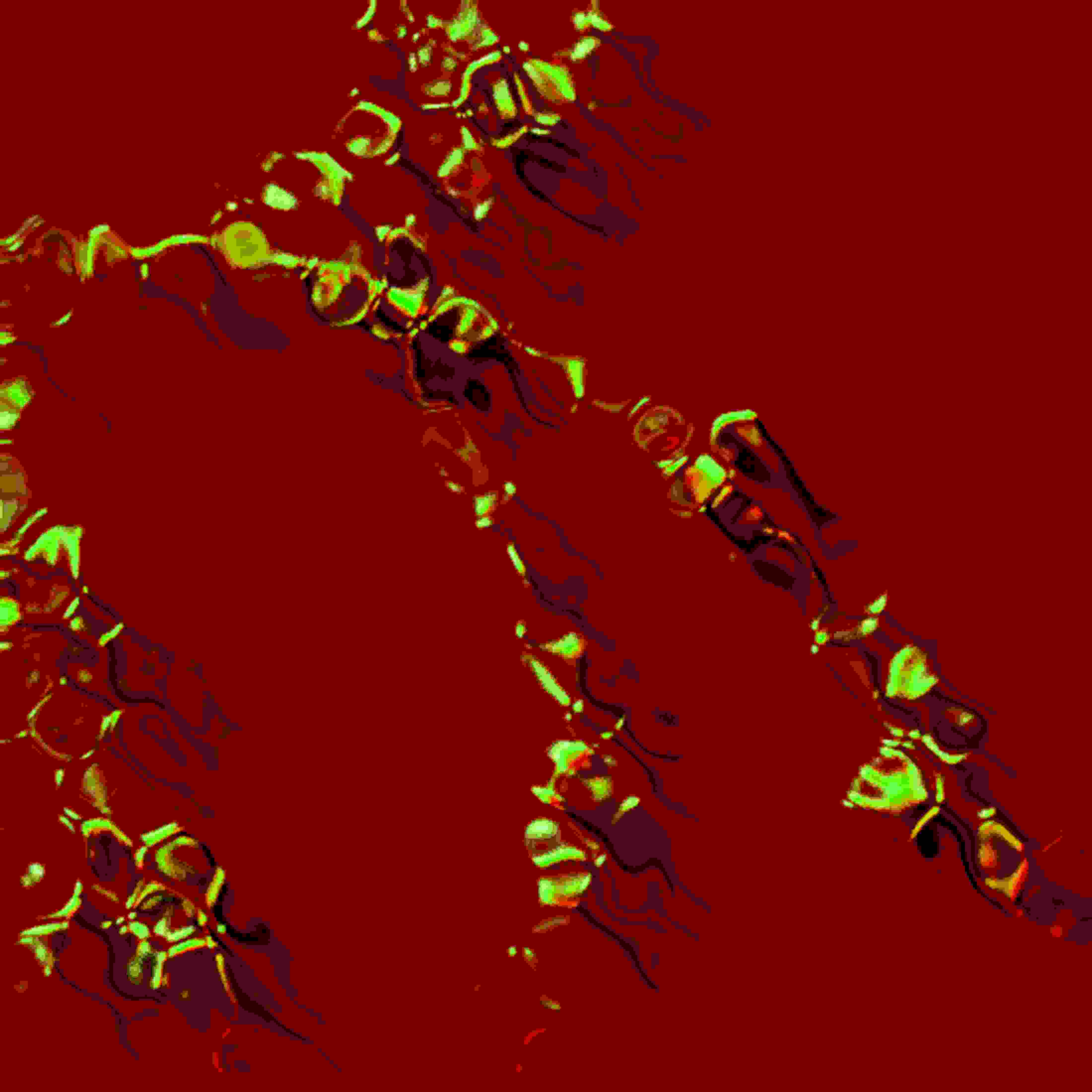
Dr.Eduard Korkotian Weizmann Institute of Science Rehovot, ISRAEL The Dying Neuron The image is a 3D reconstructed piece of dendritic tree of an apoptotic hippocampal neuron in culture. The shading is produced using home-made Matlab software. |
|
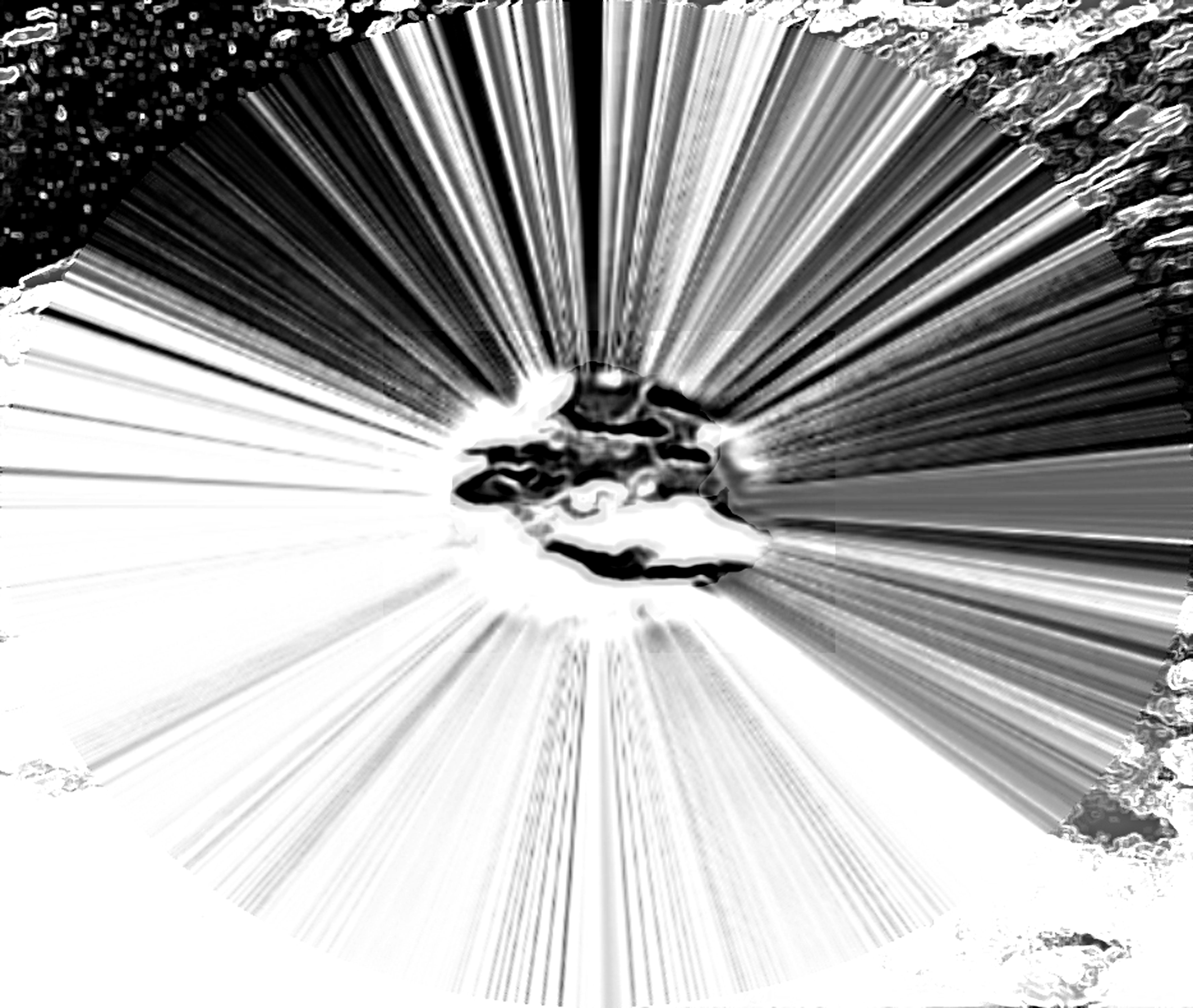
Dr.Eduard Korkotian Weizmann Institute of Science Rehovot, ISRAEL Energy of Light The image is a micro-hole produced by thin Ultra Violet 4 nanosecond lasting strong laser flash in biological structure. The micro-hole diameter is about 1 micron. Such laser flashes are used for different types of micro-stimulations and fine manipulations. Image is additionally processed using Adobe Photoshop software. |
|
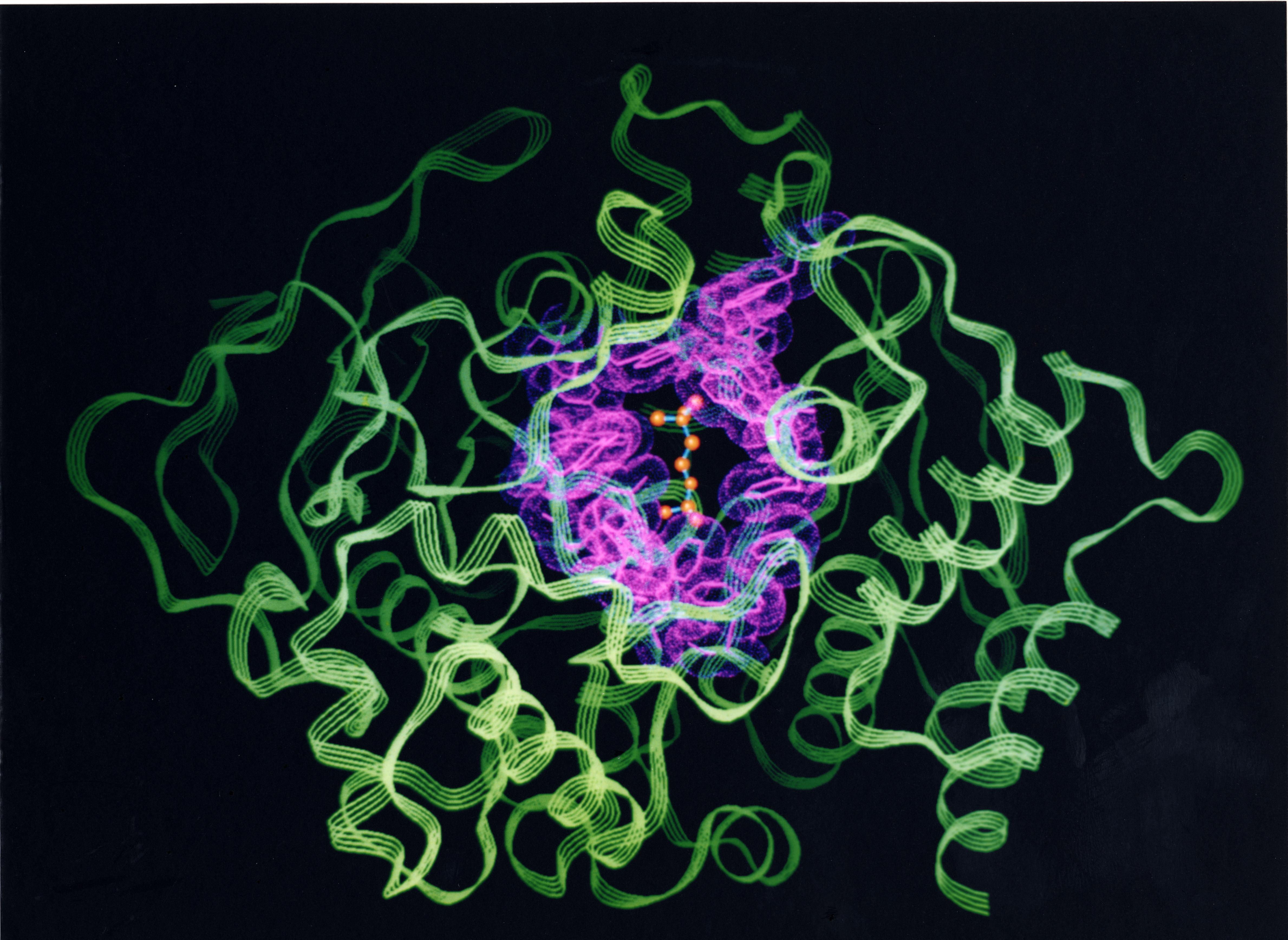
| Prof.Joel Sussman Weizmann Institute of Science Rehovot, ISRAEL Structure of T. californica Acetylcholinesterase. 3D ribbon trace of the structure of T. californica acetylcholinesterase, showing the 14 conserved aromatic residues that line the entrance of the active-site gorge in pink, and the model of acetylcholine in ball in stick. |
|
| Prof.Joel Sussman Weizmann Institute of Science Rehovot, ISRAEL Acetylcholinesterase Acetylcholinesterase (AChE) shown as a surface in yellow, with the snake toxin inhibitor fasciculin-II shown in green. The red surface corresponds to the footprint of fasciculin-II on AChE, and the white region the active-site gorge. (Image made together with Dr. Gitay Kryger) |
|
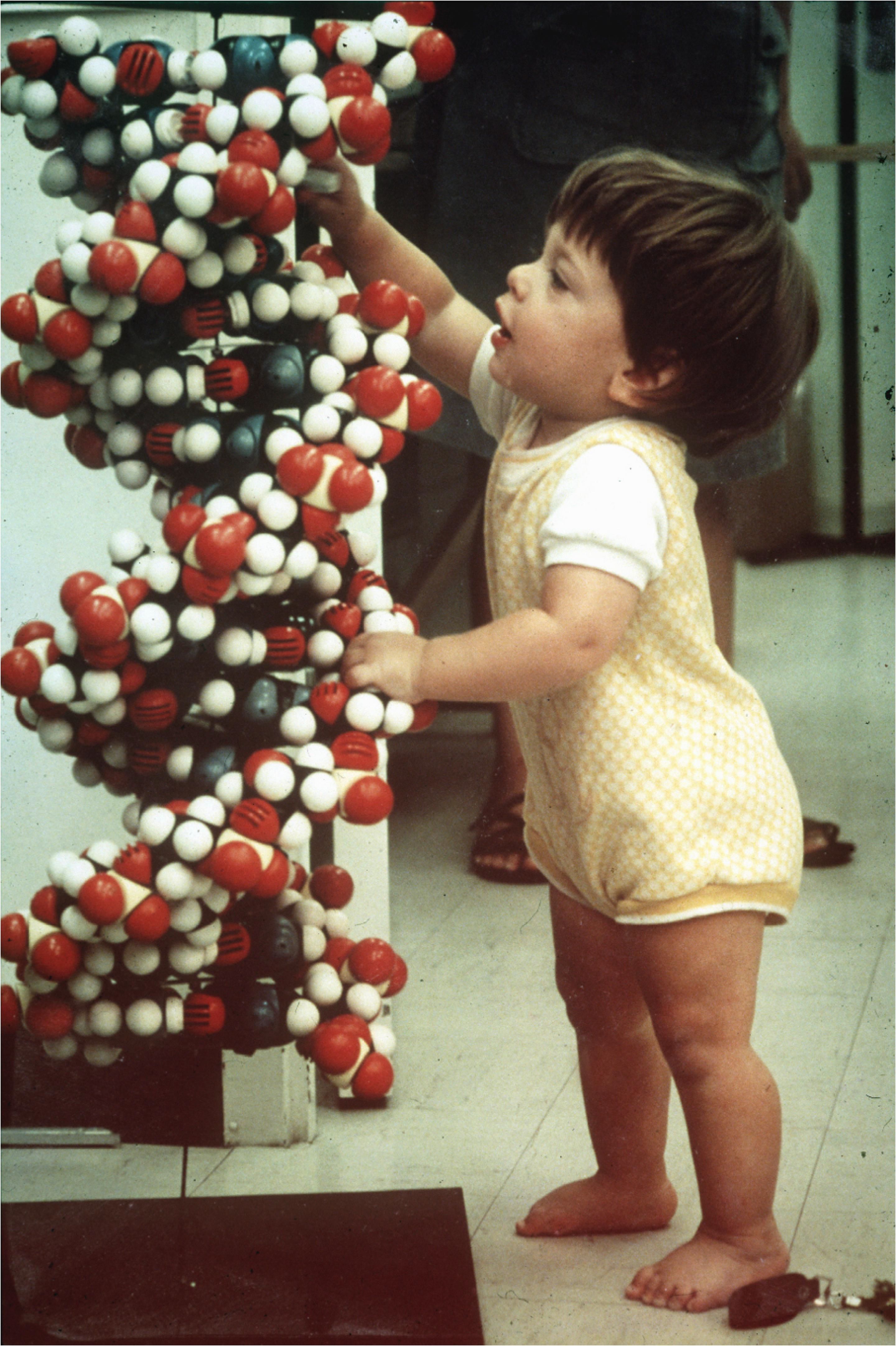
Prof.Joel Sussman Weizmann Institute of Science Rehovot, ISRAEL Photo of Ehud Z Sussman (at 2 years old) examining a 3D plastic model of DNA. (Photo taken by Prof. Sung-Hou Kim). |

|
Prof.Joel Sussman Weizmann Institute of Science Rehovot, ISRAEL Acetylcholinesterase. A cross section through the active site gorge of acetylcholinesterase. The molecular surface is shown in lavender, and electrostatic field vectors in green. The catalytic triad, S200, E327, and H440 are shown in X,Y, and Z, respectively. W279 at the top of the gorge, and W84, adjacent to the triad, are shown in white. |
PDF Available |
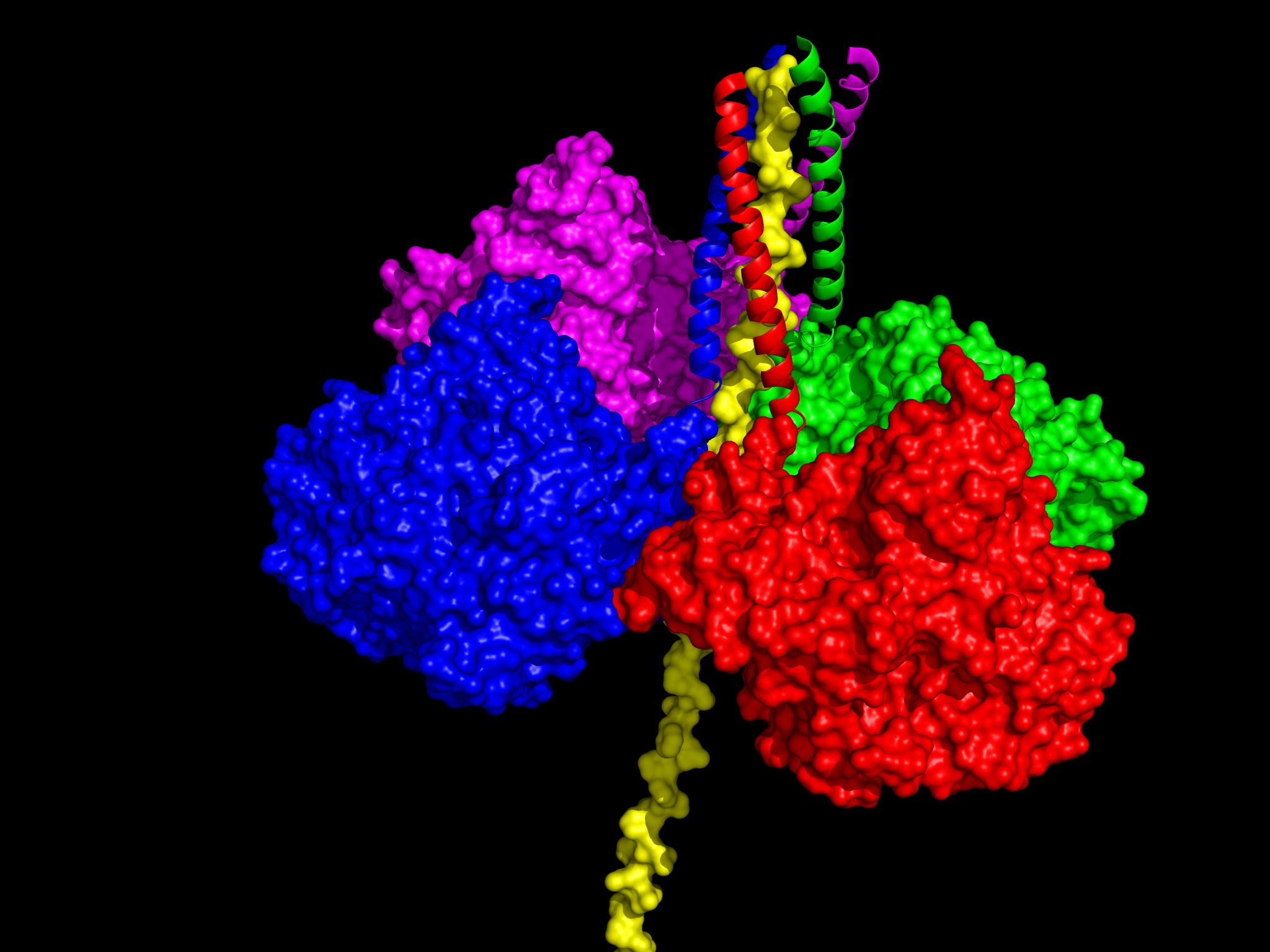
Prof.Joel Sussman Weizmann Institute of Science Rehovot, ISRAEL Model of the physiological Acetylcholinesterase tetramer. View in which the ColQ polypeptide is vertical. The WAT polypeptides are displayed as ribbons, and the PRAD as a yellow surface model. Each of the four WATs (magenta, red, blue and cyan) assumes a coiled-coil conformation, together forming a left-handed superhelical structure. |
|
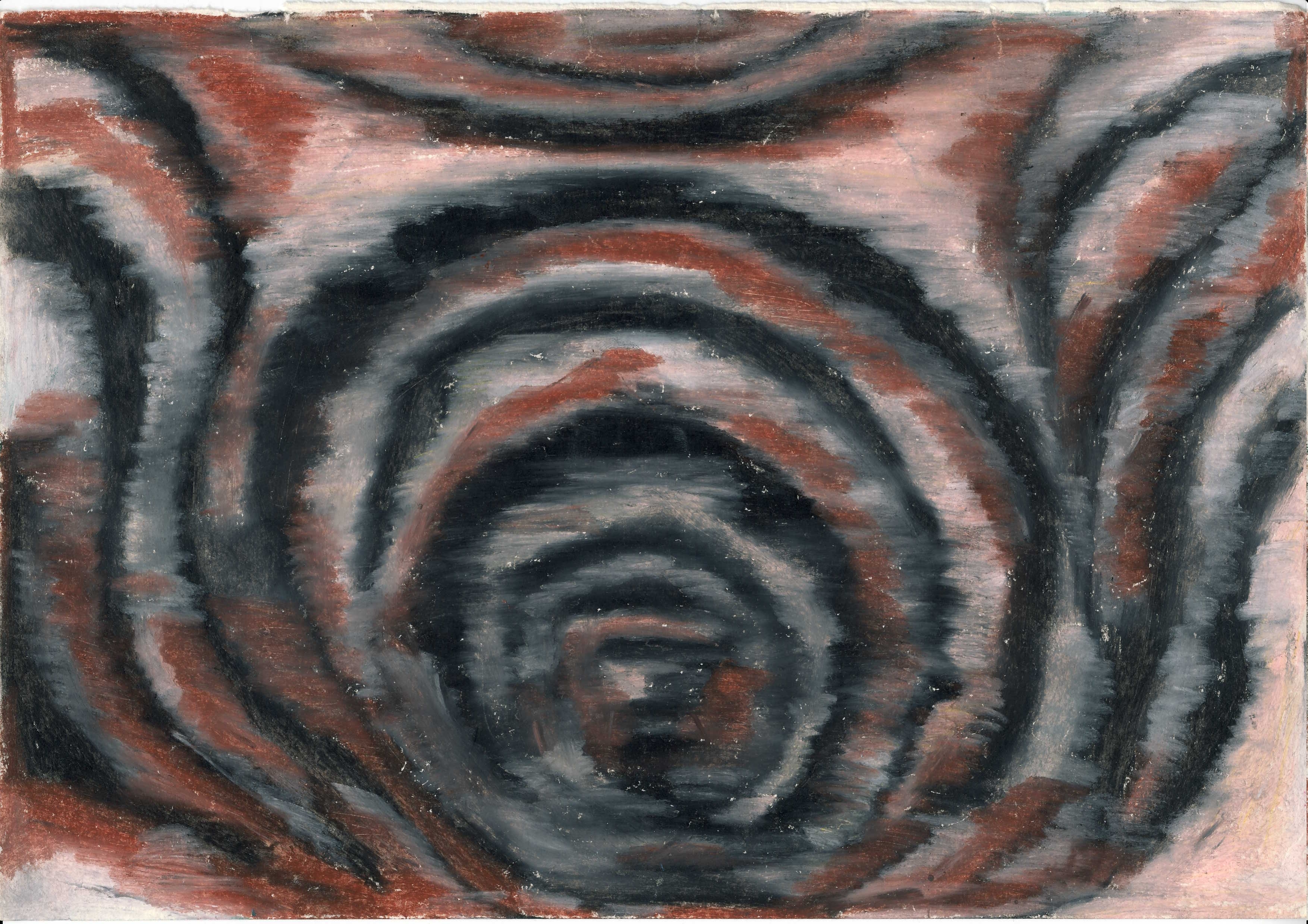
Dr. Milana Frenkel-Morgenstern Weizmann Institute of Science Rehovot, ISRAEL Light Reflection on Plastic Window. Oil Crayola. 1993. |
|
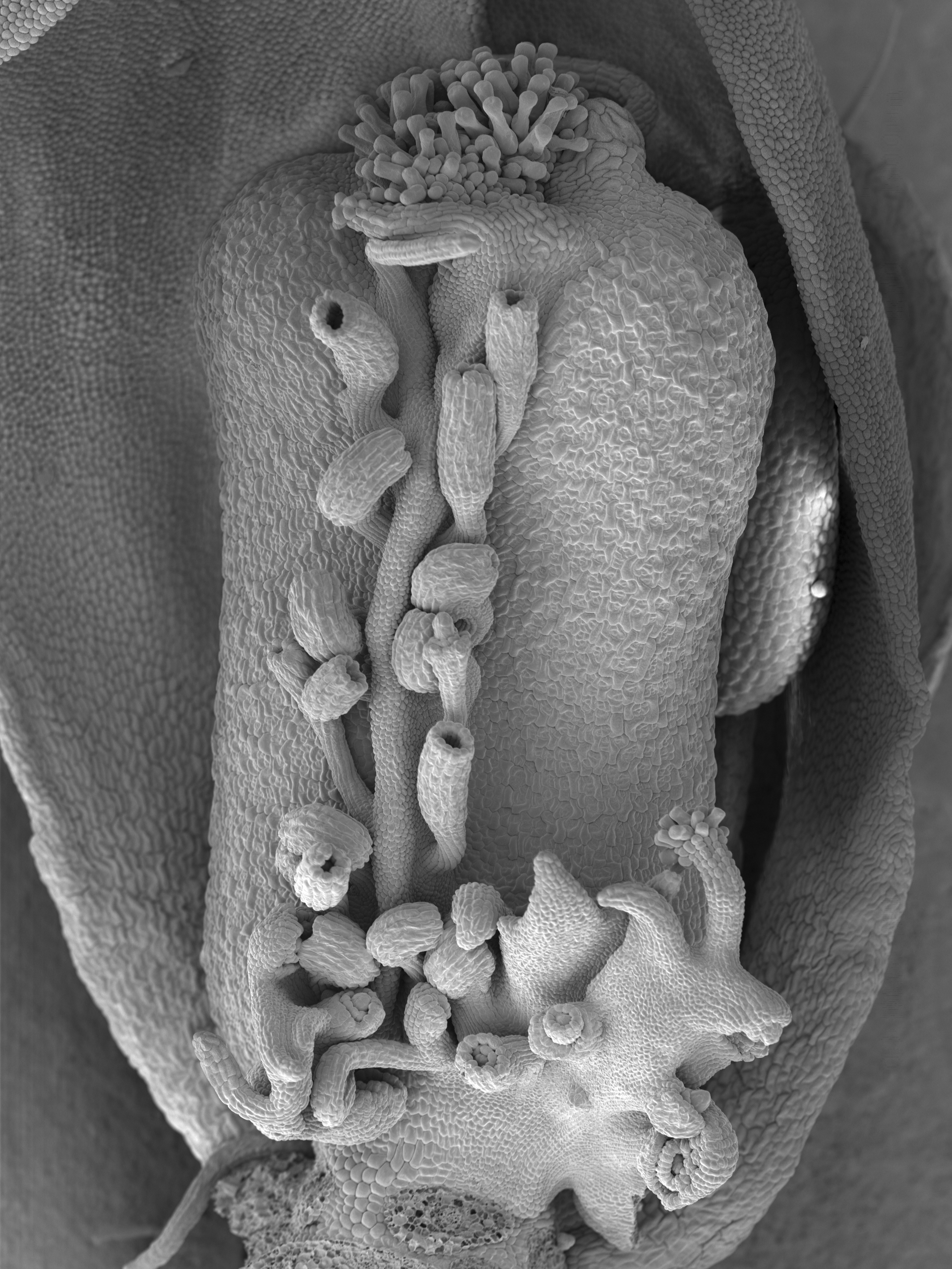
Prof.Yuval Eshed Weizmann Institute of Science Rehovot, ISRAEL Arabidopsis carpels. Carpels, the female organs of the plant, protect the seeds and will become the mature fruit. However, specific mutations can simulate formation of internal organs at the outer side of young carpels. In extreme cases, this can result in seeds developing inside and outside of the fruit. |
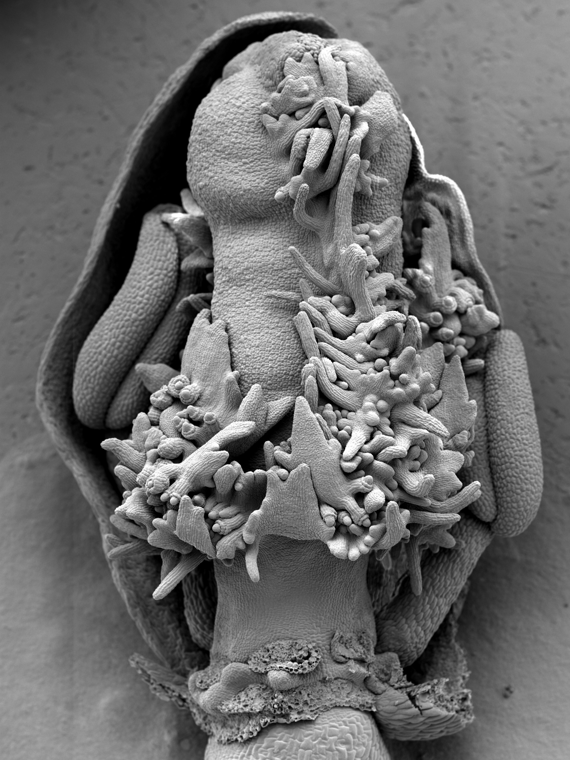
|
Prof.Yuval Eshed Weizmann Institute of Science Rehovot, ISRAEL Arabidopsis carpels. Carpels, the female organs of the plant, protect the seeds and will become the mature fruit. However, specific mutations can simulate formation of internal organs at the outer side of young carpels. In extreme cases, this can result in seeds developing inside and outside of the fruit. |
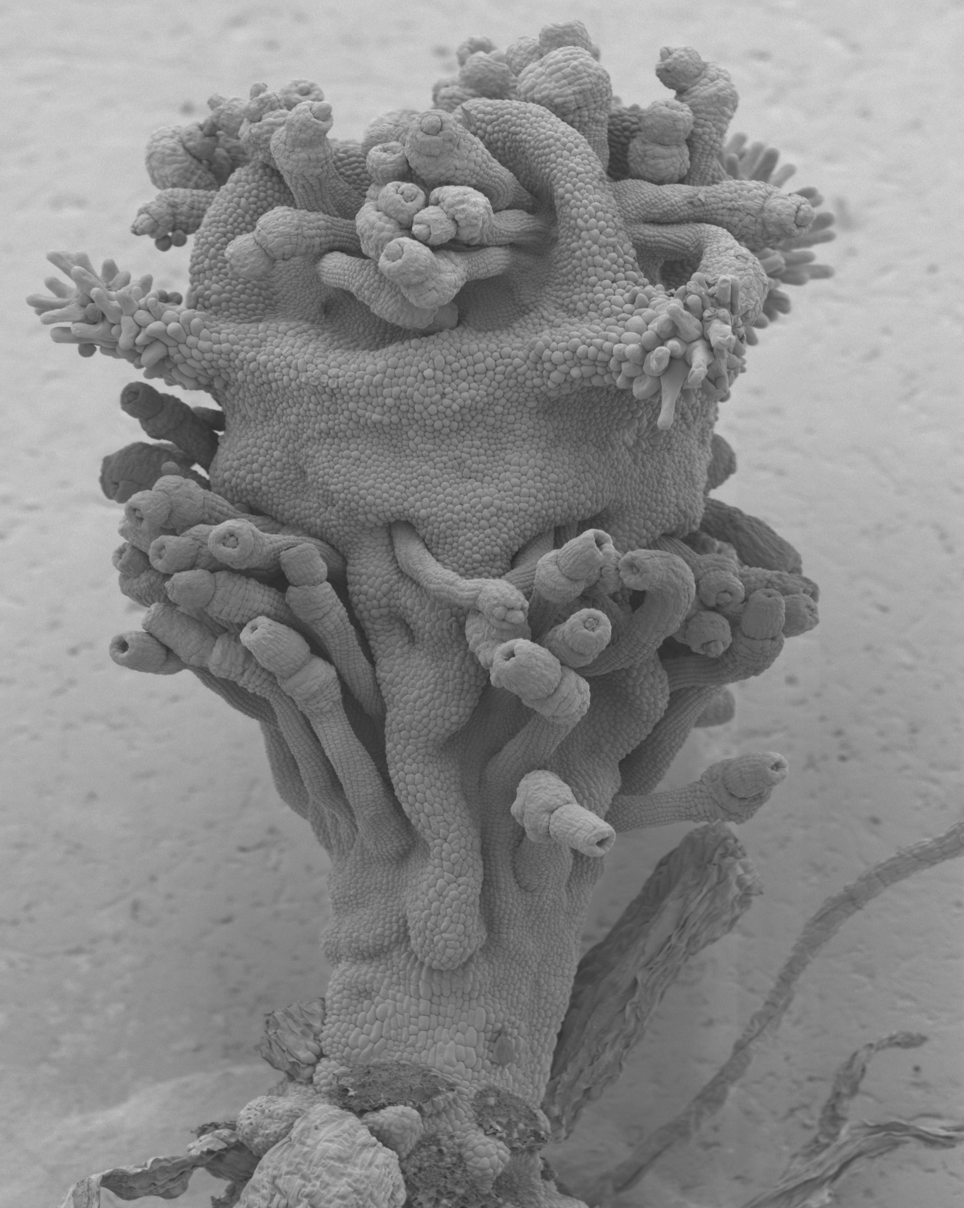
|
Prof.Yuval Eshed Weizmann Institute of Science Rehovot, ISRAEL Arabidopsis carpels. Carpels, the female organs of the plant, protect the seeds and will become the mature fruit. However, specific mutations can simulate formation of internal organs at the outer side of young carpels. In extreme cases, this can result in seeds developing inside and outside of the fruit. |
|
|
Mr. Benjamin Jefferys Imperial College London, UK Deliberately obfuscated visualisation of forces hypothesized to be important for protein folding in a TIM barrel domain This is a view of some of the forces which we suspect are important in protein folding in a model developed in our lab. Hollow tubes are rendered which represent the forces between sidechains and backbone of a simplified model of a TIM barrel domain. The tubes initially change size and length as the points the forces operate between move to their satisfaction. The view has been deliberately exaggerated to present a chaotic and incomprehensible view of protein structure and dynamics which is distinct from the artificially simplified and structurally biased view of proteins we are familiar with. It might be seen as a representation of the complexity of the protein folding problem which has vexed so many eminent researchers over the decades. It is a reminder that the simplifications we make to the biological systems we study have a profound influence on our perception of those problems. It is also a demonstration of the beauty inherent in a complex system, even when we don't fully understand it. | Video Available |
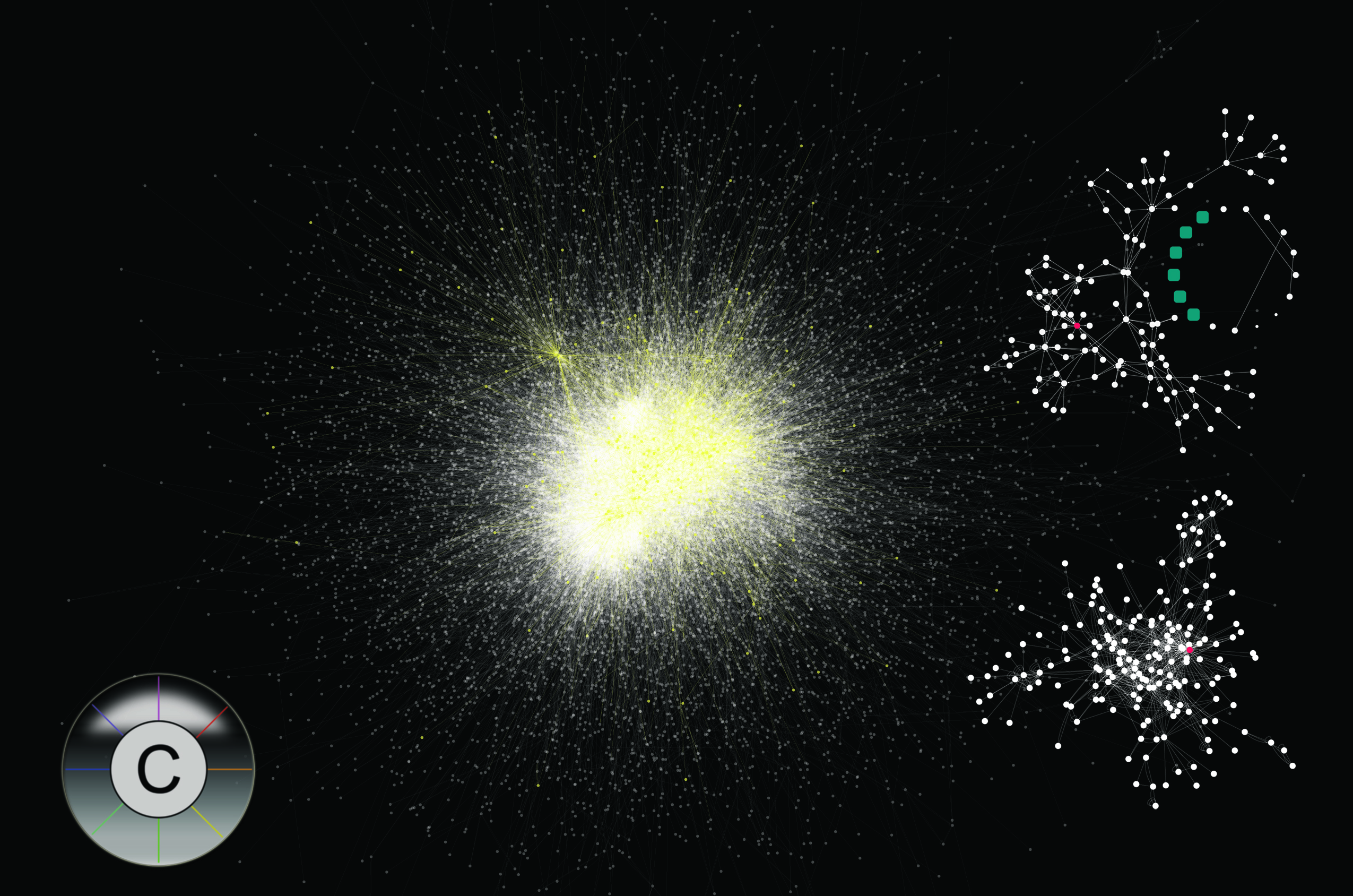
Mr. Keiichiro Ono University of California, San Diego, USA Human Interactome Analysis of biological networks is one of a hot topic in biology today. To understand the complex biological circuits (i.e., protein-protein interactions, protein-DNA interactions, etc.) data integration and visualization are two important technology. This image represents a typical workflow of biologists working on this hard problem. The center image is known human protein-protein interactions (called interactome) visualized by Cytoscape (http://www.cytoscape.org). This network is created by merging multiple data sources, including HPRD, BIND, BioGRID, and IntAct. Yellow nodes are proteins exist on known pathway (MAPK Signaling Pathway in KEGG database) and lower-right image is a visualization of the sub network. Upper-right network image is a visualization of the MAPK Signaling Pathway directly taken from KEGG database. You may notice that both of the smaller images have a red node. It is MAPK14 protein (Entrez Gene ID 1432). This type of visualization is useful when you want to map your own experimental data onto known pathways. All of these data integration, mapping, and visualization were done in Cytoscape. |
 |
Mr. Jared Flatow Northwestern University, USA A Portrait of James D. Watson in his own Words According to Wikipedia, "a portrait is a painting, photograph, sculpture, or other artistic representation of a person". Instead of simply using color pigments, we use unique portions of Dr. James Watson's DNA sequence to portrait himself. Dr. Watson was the discoverer of the structure of the DNA and helped to establish the Human Genome Project. DNA, as a primary genetic material, defines the molecular signature of oneself. Dr. Watson's DNA was fully sequenced and made public in 2007 by The Baylor College of Medicine Genome Sequencing Center, 454 Life Sciences Technology, and The Rothberg Institute. We used the SNPs, which define the small differences of DNA from person to person, to uniquely represent Dr. Watson. In order to do this, we took the variant allele base pairs from Dr. Watson's genome (Cold Spring Harbor Laboratory distribution, 6/6/2007) which had a sequence observation count greater than 12, and generated a portrait capturing his phenotype. |
PDF Available |
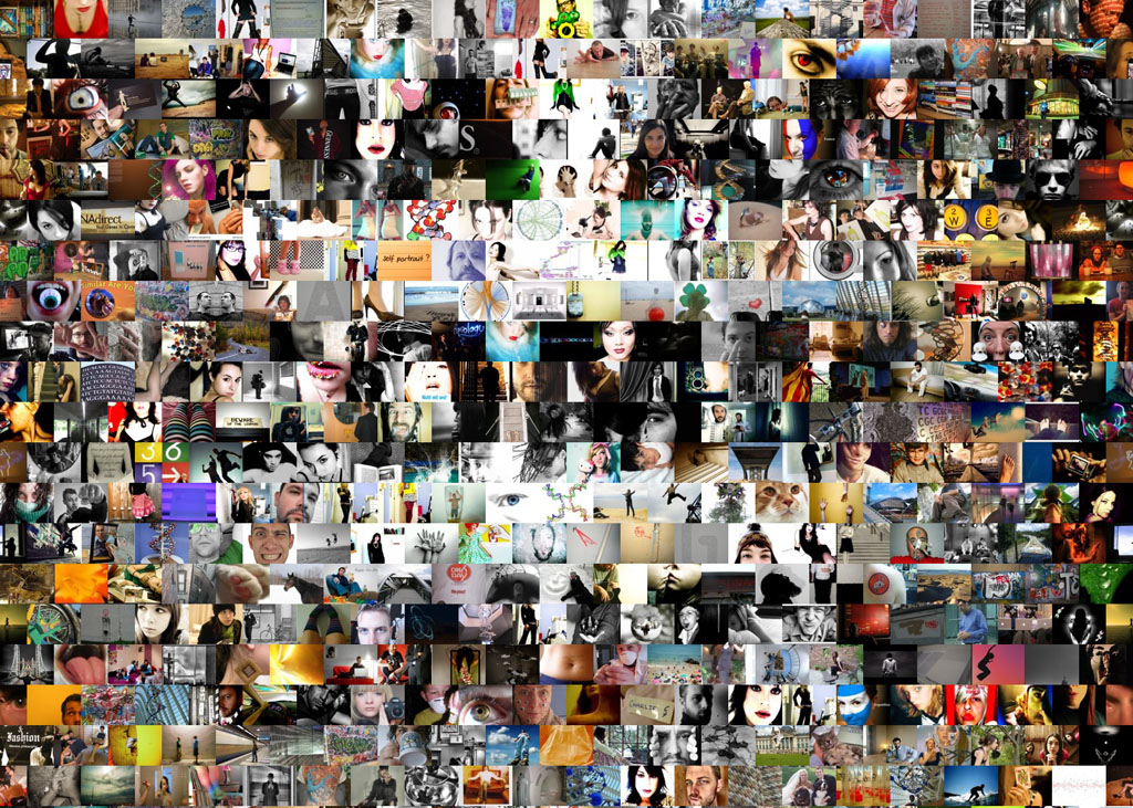
| Dr. Simon Lin Northwestern University, USA DNA and Community Artists constantly explore the interactions between science and society. We looked into the public understanding of DNA in the Web 2.0 era by retrieving Creative Commons (CC)-licensed photos from the Flickr (photo-sharing) website. We retrieved 899 images using the topics of DNA and myself on April 6, 2008. We rearranged these images using a mosaic algorithm to reveal the hidden message of "DNA and Community". Traditional art uses oil and brush; we are using Python and the internet to experiment with new building blocks of CC-licensed photos. By integrating the photos through the lens of 899 individuals, we are investigating how people share their life stories (Flickr) and how people share their creative responsibility (CC license). It is interesting to note that our work is also licensed under CC and thus has 899 lines of acknowledgements. |
 |
|
Dr. Peter D Karp SRI International, AE206, USA EcoCyc E. coli Metabolic Network The artwork is a depiction of the Escherichia coli metabolic network and transporter complemented that was generated automatically from the EcoCyc database by the Pathway Tools software. |
PDF Available |
|
Dr. Kazuharu Arakawa Institute for Advanced Biosciences, Keio University, Japan E-Cell 3D: 3-Dimensional Visualization of Dynamic Cell Simulation Since 1996, E-Cell Project in Institute for Advanced Biosciences has been aiming to model and reconstruct biological phenomena in silico, and developing necessary theoretical supports, technologies and software platforms to allow precise whole cell simulation. Cell simulation realizes theoretical experiments in silico to enhance our understanding of dynamic cellular activities through integrative systems biology approaches. E-Cell Simulation version 3 (E-Cell SE 3) is a potential platform for this purpose, which allows object-oriented simulations with multiple algorithms and timescales, with scriptable front-end using Python programming language. E-Cell 3D is a novel interface for the system to aid the understanding of highly complex systems by researchers in the process of modeling through intuitive visualization using 3D graphics. The software and documentations are freely available at http://ecell3d.iab.keio.ac.jp. |
PDF Available and Video Available |
|
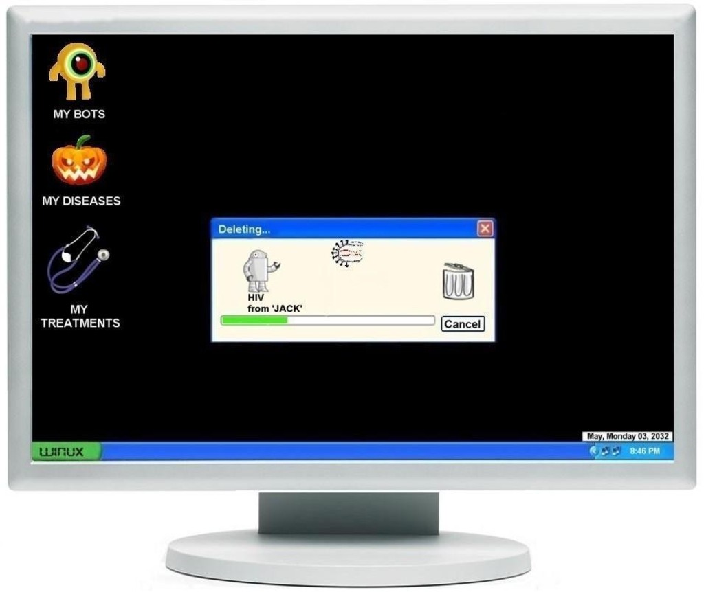
Mr. Simarjotsingh P Pabla Sardar Patel University, India BIological Operating System- BIOS "For tomorrow belongs to the people who prepare for it today" - African Proverb In future, advances in bioinformatics, bioengineering and nanotechnology will result in development of a comprehensive "operating system". These kinds of software packages would deal with wide array of health issues at the "molecular plane". The work force behind prognosis, diagnosis and treatments would be NANOBOTS. Bots will communicate with the BIOS via specific hardware and orchestrate the treatment. Initially its availability will be constrained to experts only. BIOS would be able to download new treatments as well as updates for new diseases from the internet, for example downloading new HIV drug resistant strain information. Further augment in technology would result in portability of BIOS to devices like iPODs and PSPs. Perhaps, BIOS would be the path breaking achievement that would bring forth the best of molecular biology, computational biology, bioengineering and nanotechnology. |
 |
|
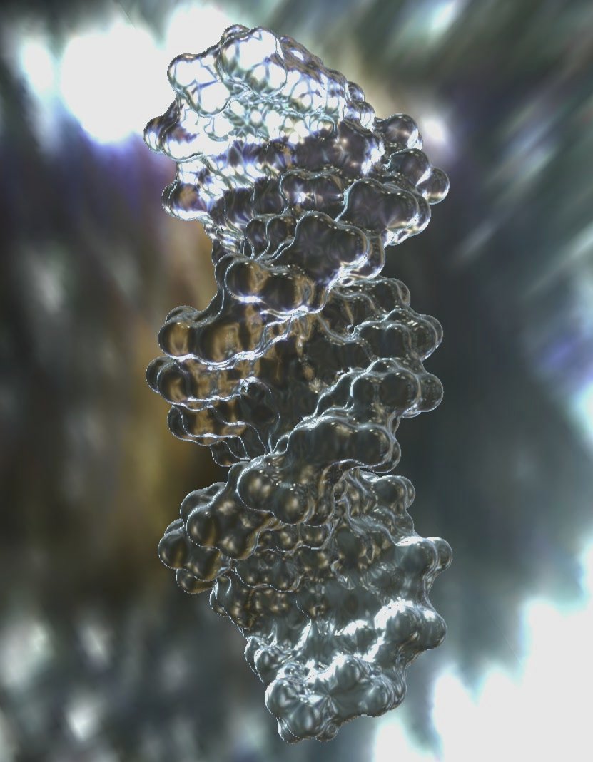
Mr. Matthieu Chavent CNRS, Loria, France High Quality Visualization of Molecular Skin Surface The Solvent Excluded Surface (SES) or Molecular Surface (MS) is the most widely-used surface for representing macromolecular assemblies. Starting from the pioneering algorithm proposed by Connolly, numerous works have been devoted to the improvement of the related methods, in order to provide fast and robust generation of high quality pictures of MS. However, existing software discretize the surface with triangles or grids so that a level of zoom is always found where triangles or cubes appear. Furthermore, the generated MS is not exempt from singularities due to self intersections. With our program: MetaMol, we tackle the problems of precision and singularities by using the Skin Surface, as defined by H. Edelsbrunner at the end of the 90's. This particular surface definition is referred to as the Molecular Skin Surface (MSS) when applied to molecular assemblies. It is basically comparable to the MS representation but providing additional smoothness and decomposability. Another advantage of the MSS is that the nature of the surface depends on a single parameter, the shrink factor. Adjusting it allows to evolve in real time from a van der Waals surface to the MSS (closed to the MS) until a simplified surface that can be very useful for coarse-grain protein docking. We use a ray-casting method implemented directly on GPU. This has two advantages: the ray-casting algorithm directly uses the MSS equation and does not need to resample it and pixel-accurate images are generated. Furthermore, MetaMol provides nice lighting effects that enhance the displaying quality. |
Video Available |
Ellen K.Levy Visiting Scholar NYU and PhD candidate at Z-Node, an interdisciplinary doctoral program for artists involved with science and technology. Proliferation 2 (Mad Cows). 2001 |

|
|
Mallika Veeramalai Burnham Institute for Medical Research, La Jolla, US. TOPSAN (The Open Protein Structure Annotation Network) The Open Protein Structure Annotation Network (TOPSAN) is a wiki designed to collect, share and distribute information about protein three-dimensional structures, and to advance it towards knowledge about functions and roles of these proteins in their respective organisms. TOPSAN will serve as a portal for the scientific community to learn about protein structures and also to contribute their expertise in annotating protein function. Access and contribute to the TOPSAN via https://www.topsan.org/WikiHome. |

|