


Award Winners - ISMB/ECCB 2015
Ian Lawson Van Touch Memorial Award
Springer Outstanding Poster Award
TP025: Identification of causal genes for complex traits
Presenting Author: Farhad Hormozdiari, University of California, Los Angeles, United States
Additional authors:
Gleb Kichaev, University of California, Los Angeles, United States
Wen-Yun Yang, University of California, Los Angeles, United States
Bogdan Pasaniuc, University of California, Los Angeles, United States
Eleazar Eskin, University of California, Los Angeles, United States
Motivation: Although genome-wide association studies (GWAS) have identified thousands of variants associated with common diseases and complex traits, only a handful of these variants are validated to be causal. We consider “causal variants” as variants which are responsible for the association signal at a locus. As opposed to association studies that benefit from linkage disequilibrium (LD), the main challenge in identifying causal variants at associated loci lies in distinguishing among the many closely correlated variants due to LD. This is particularly important for model organisms such as inbred mice, where LD extends much further than in human populations, resulting in large stretches of the genome with significantly associated variants. Furthermore, these model organisms are highly structured, and require correction for population structure to remove potential spurious associations.
Results: In this work, we propose CAVIAR-Gene, a novel method that is able to operate across large LD regions of the genome while also correcting for population structure. A key feature of our approach is that it provides as output a minimally sized set of genes that captures the genes which harbor causal variants with probability . Through extensive simulations, we demonstrate that our method not only speeds up computation, but also have an average of 10% higher recall rate compared to the existing approaches. We validate our method using a real mouse high-density lipoprotein data (HDL) and show that CAVIAR-Gene is able to identify Apoa2 (a gene known to harbor causal variants for HDL), while reducing the number of genes that need to be tested for functionality by a factor of 2.
The software is freely available for download at genetics.cs.University of California, Los Angeles.edu/caviar
OP12: Pathway relevance ranking for tumor samples through network-based data integration
Presenting Author: Lieven Verbeke, Ghent University / iMinds / IBCN, Belgium
Abstract:
We present a new pathway relevance ranking method that is able to prioritize pathways according to the information contained in any combination of tumor related omics datasets. Key to the method is the conversion of all available data into a single network representation containing not only genes but also individual patient samples. Additionally, all data are linked through a network of previously identified molecular interactions. The performance of the new method is demonstrated by applying it to breast and ovarian cancer datasets from The Cancer Genome Atlas. By integrating gene expression, copy number, mutation and methylation data, the method’s potential to identify key pathways involved in breast cancer development shared by different molecular subtypes, is illustrated. Interestingly, certain pathways were ranked equally important for different subtypes, even when the underlying (epi)-genetic disturbances were diverse. The pathway ranking method was also able to identify subtype-specific pathways. Often the score of a pathway could only be explained by a combination of genetic and epi-genetic disturbances, stressing the need for a network-based data-integration approach. The analysis of ovarian tumors, as a function of survival-based subtypes, demonstrated the method’s ability to correctly identify key pathways, irrespective of tumor subtype. A differential analysis of survival-based subtypes revealed several pathways with higher importance for the bad-outcome patient group than for the good-outcome patient group. Many of the pathways exhibiting higher importance for the bad-outcome patient group could be related to ovarian tumor proliferation and survival.
OP14: Novel brain-specific miRNA discovery using small RNA sequencing in post-mortem human brain
Abstract:
MicroRNAs (miRNA) are short non-coding RNAs that regulate gene expression mainly through translational repression of target mRNA molecules. More than 2700 human miRNAs have been identified and some are known to display tissue-specific patterns of expression. Here, we use high-throughput small RNA sequencing to discover novel and possibly brain-specific miRNAs in 94 human post-mortem prefrontal cortex samples from patients with Huntington's disease and Parkinson's disease and normal neuropathology. Using a custom analysis pipeline, we identified 66 novel miRNA candidates that originate in both intergenic and intragenic regions of the genome. 21 of the candidate miRNAs show sequence similarity with known mature miRNA sequences and may be novel members of known miRNA families, while the remaining 45 may constitute previously undiscovered families of miRNAs that are specific to the brain. In a small number of these novel miRNAs, preliminary differential expression analysis between neurodegenerative disease and normal samples identified differences in expression. These results suggest that a portion of these novel miRNAs may not only be unique to brain, but may have a role in the neurodegenerative disease processes.
OP15: Computationally efficient approach for novel transcript discovery across large RNA-seq dataset reveals glioblastoma-associated lncRNAs
Presenting Author: Maria Laaksonen, BioMediTech, University of Tampere, Finland
Additional Authors:
Presenting Author: Helen McCormick, Victor Chang Cardiac Research Institute, Australia
OP30: Determining the winning SH3 coalition: how cooperative game theory reveals the importance of domain residues in peptide binding
Presenting Author: Ashley Conard, United States
OP13: ContiBAIT: An R Package for Genome Finishing Using Strand-seq
Presenting Author: Kieran O’Neill, British Columbia Cancer Agency, Canada
OP16: Tau Protein Related Acetylation of Histone 3 Lysine 9 in the Human Brain
Presenting Author: Hans-Ulrich Klein, Harvard Medical School, United States
Additional Authors:
A&S11 Analogue Alignment
 “Multiple sequence alignments were once performed manually, and even today, we still examine automatically computed alignments to check that we can't do better.” –Jim Procter
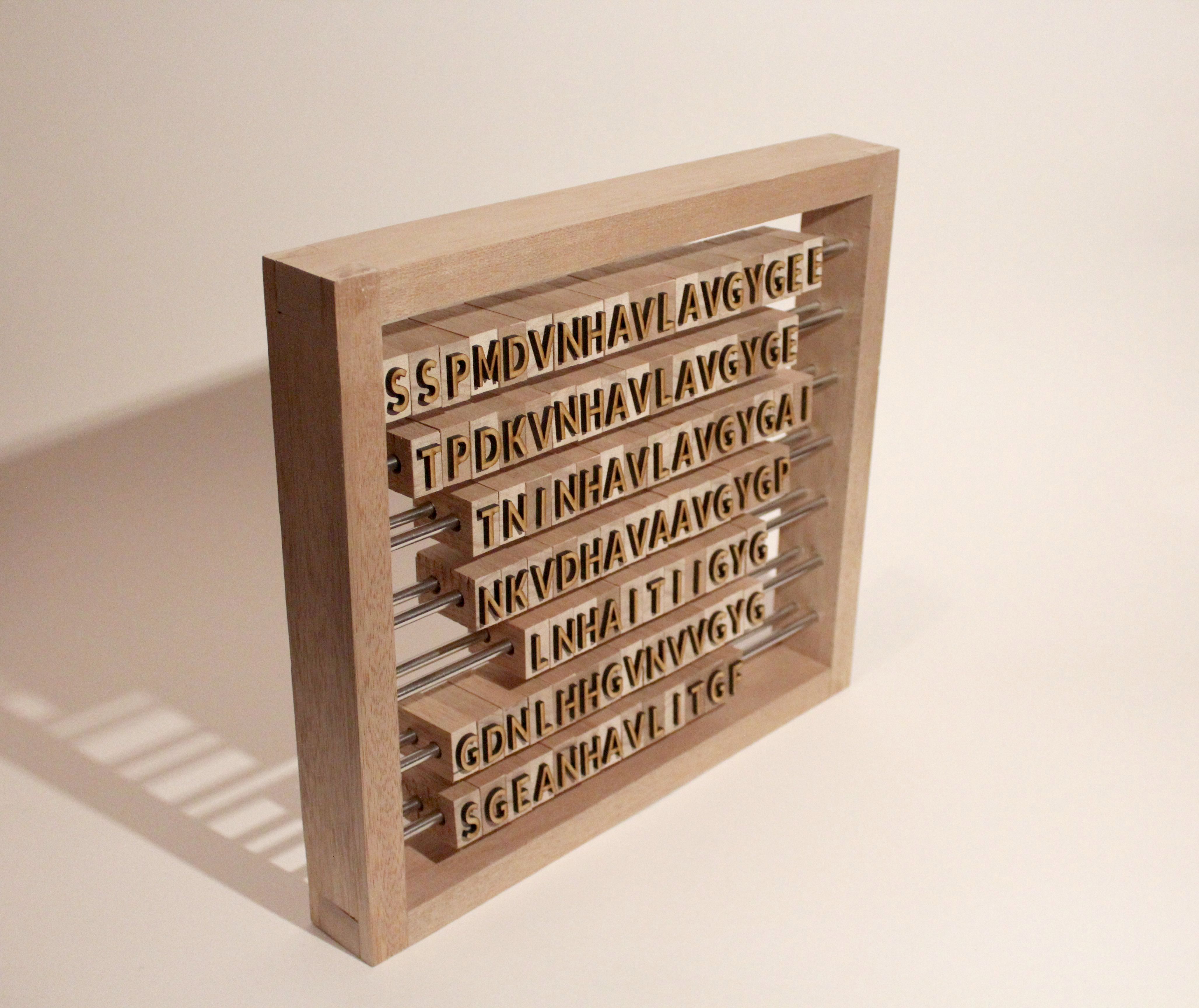
This is an image of the Jalview Abacus, a sculptural attempt to visually represent the function of the Jalview protein alignment program. The program can be used to find alignments of amino acids in similar proteins. These alignments are then used to find similarities and differences between these proteins.
“Multiple sequence alignments were once performed manually, and even today, we still examine automatically computed alignments to check that we can't do better.” –Jim Procter
This is an image of the Jalview Abacus, a sculptural attempt to visually represent the function of the Jalview protein alignment program. The program can be used to find alignments of amino acids in similar proteins. These alignments are then used to find similarities and differences between these proteins.
This object expresses the core process of Jalview in a physical space, and plays on the relationship between high tech and low tech solutions. It is a functioning abacus built by hand from wood and steel. Each row is an extract from different similar proteins (cysteine proteases) and an alignment can be found by lining up the beads of like amino acids in the columns. If it was long enough it could be used to align the entire sequence manually.
Photography: Luke Wilson
Design and Construction: Luke Wilson in collaboration w. Jim Procter and Geoff Barton
Program Menu
ISCB Board of Directors
Alfonso Valencia, ISCB President, Spanish National Cancer Research Centre (CNIO), Spain
Burkhard Rost, ISCB Past President, Technical University Munich, Germany
Bonnie Berger, ISCB Vice President, MIT, United States
Terry Gaasterland, ISCB Vice President, University of California San Diego, United States
Thomas Lengauer, ISCB Vice President, Max-Planck Institute for Informatics, Germany
Christine Orengo, ISCB Vice President, University College London, United Kingdom
Reinhard Schneider, ISCB Treasurer; University of Luxembourg, Luxembourg
Scott Markel, ISCB Secretary, Dassault Systèmes BIOVIA, United States
DIRECTORS
Teresa Attwood, University of Manchester, United Kingdom
Alex Bateman, European Bioinformatics Institute, United Kingdom
Judith A. Blake, The Jackson Laboratory, United States
Alan Christoffels, University of the Western Cape, South Africa
Manuel Corpas, The Genome Analysis Centre, United Kingdom
Bruno Gaëta, The University of New South Wales, Australia
Paul Horton, AIST, Japan
Jigisha Anupama, University College Dublin, Ireland
Janet Kelso, Max Planck Institute for Evolutionary Anthropology, Germany
Richard H. Lathrop, University of California Irvine, United States
Fran Lewitter, Whitehead Institute, United States
Michal Linial, The Hebrew University of Jerusalem, Israel
Francisco Melo Ledermann, Pontificia Universidad Catolica de Chile
Jill Mesirov, The Broad Institute of MIT and Harvard, United States
Nicola Mulder, University of Cape Town, South Africa
Predrag Radivojac, Indiana University, United States
Anna Tramontano, University of Rome "La Sapienza", Italy
Olga Troyanskaya, Princeton University, United States
Lonnie Welch, Ohio University, United States
EX OFFICIO DIRECTOR
Martin Vingron, Max Planck Institute for Molecular Genetics, Germany
ECCB Committee Members
Ron Appel, Swiss Institute of Bioinformatics, Switzerland
Søren Brunak, Technical University of Denmark and University of Copenhagen, Denmark
Marie-Dominique Devignes, CNRS, University of Lorraine, Nancy, France
Jaap Heringa, Vrije Universiteit, Amsterdam, The Netherlands
Oliver Kohlbacher, University of Tübingen, Germany
Michael Linial, The Hebrew University of Jerusalem, Israel
Rodrigo Lopez, European Bioinformatics Institute, EMBL-EBI, Cambridge, United Kingdom
Yves Moreau, KU Leuven, Belgium
Marie-France Sagot, Inria Grenoble Rhône-Alpes, Lyon, France
Torsten Schwede, SIB Swiss Institute of Bioinformatics & Biozentrum, University of Basel, Switzerland
Janet Thornton, European Bioinformatics Institute, Hinxton, United Kingdom
Anna Tramontano, Sapienza University of Rome, Italy
Alfonso Valencia, Spanish National Cancer Research Centre (CNIO), Madrid, Spain
Jaak Vilo, University of Tartu, Estonia
Martin Vingron, Max Planck Institute for Molecular Genetics, Berlin, Germany
General Information - ISMB 2016
- About Us
- Accomodation
- Awards
- Airport Shuttle
- Code of Conduct
- Child Care
- Disney World
- Universal Studios
- Volunteer
Special Talks - ISMB 2014
Room: TBA
Date/Time: Sunday, July 13 at 11:30 a.m. - 11:55 p.m.
The advent of the first enzyme structures in the 1960s, coupled to increasing computer power at the time, marked a turning point for computational enzymology. Specifically, starting in 1970, a number of different QM+MM and QM/MM approaches were introduced by Warshel and coworkers to facilitate the description of reactions in enzymes. This and molecular dynamics simulations of biological reactions (that also started with Warshel’s work), as well as the development of classical force fields, mark the emergence of multiscale models for chemical reactivity, that allowed us to begin to directly translate structural information into an energetic picture, to better understand enzyme function. In my view the most effective direction to address this problem has been the Warshel’s 1980s “empirical valence bond” approach. Despite its seemingly theoretical simplicity, the empirical valence bond approach remains one of the most powerful tools to understand chemical reactivity in biological systems even today. This talk will explore the theoretical basis and historical background for this approach, and illustrate its application to a number of the most challenging problems in computational enzymology. Additionally, the unimaginable gains in computational power of recent decades have allowed for ever more complex systems to be addressed. Therefore, this talk will conclude by discussing the power of the EVB approach to address 21st Century challenges such as enzyme design, understanding protein evolution, and addressing chemical reactivity in even such big biomolecular systems as GTP hydrolysis on the ribosome.
Speaker Information
Presentation Overview
Preparing your Presentation for CCD Projectors
Speaker Ready Room
Information about the presentation computers
Presentation information for Students, Post Docs and Young Investigators!
Speakers are requested to review the conference schedules available on the conference website. Please note that minor schedule changes may continue to be made. Schedules are available at: https://www.iscb.org/cms_addon/conferences/ismbeccb2015/schedule/schedule.php
All parallel sessions are 20 minutes in length and there are three (3) per block. Speakers are asked to be available at the presentation room 10-15 minutes before the start of the first presentation.
Speakers should prepare a 15-minute presentation. The 3-5 minutes additional time will allow for movement to the podium and the opportunity to respond to one or two questions. Each room will have a presentation timer and sessions are chaired to ensure the program schedule is adhered to.
Please visit the Speaker Room at least one (1) day before your presentation. A technician will be there and available to assist you to place your presentation on the main presentation computer for transfer to the computer in your presentation room.
Delegates with presentations developed on Apple computers may use their own Apple computer but are requested to meet with the technician in advance so that details can be coordinated regarding the procedure for using the Apple computer for presentations. It is recommended that Apple computer users bring their own cable adapters to connect to the presentation projectors.
The speaker room is located in Liffey Meeting Room 4 (CCD) – you can ask the volunteer staff at the Information desk for directions. It is available to conference speakers to review their conference presentations and to transfer their presentations to the meeting room they will present in.
Speakers Room Hours
Saturday July 11, Noon – 5:00 p.m.
Sunday, July 12, 8:00 a.m. – 6:00 p.m.
Monday, July 13, 8:00 a.m. – 6:00 p.m.
Tuesday, July 14 8:00 a.m. – 6:00 p.m.
Information about the presentation computers
Toshiba Satellite Pro R50
Specification
- Non Reflective15.6 inch HD display
- Latest generation of Intel Core CPUs: Celeron / Pentium / Core i3 with latest Intel HD Graphics
- Windows 8.1
- 500GB (5400rpm/7200rom)
- 4GB Tiled Keyboard, Fast Gigabit LAN, ac a/g/n Wireless card,
- 2 x USB 3.0, 1 x USB 2.0, RGB, HDMI, 0.9mp Webcam, SD Card
- 7hr 25mins with removable Battery
- DVD-SM or no ODD options
- 2.3kg, 379.0(W)x258(D)x23.95(H)mm
Don't use "embedded fonts" in PowerPoint especially if there's video/audio or any other linked information in the presentation. Make sure that the Powerpoint file and video / audio-clips are put into the same folder.
Click here to download speaker tips
Please read these helpful tips on giving a quality talk at the conference.
As you prepare to give an oral presentation the following are some helpful tips for ensuring that both you and the international and interdisciplinary conference audience get the most out of your talk. As some talks will be recorded for viewing by our community for years to come, following these tips can also serve to make certain your best possible presentation serves you well in your future career.
Limit the number of slides to be presented.
A common mistake among presenters at all levels of experience is including too many slides for the allotted presentation time. We have all attended talks where the presenter either had to rush through or skip entire sections of slides due to having too many slides for the amount of time allotted to the talk. Worse is the presenter whose talk goes beyond the allotted time, and he or she ignores the session chair and/or session timer in order to give the full presentation detailed in the slides.
A rule of thumb is to have just one robust and informative slide for each minute of the presentation. Two or more slides per minute is sometimes possible, but this typically only works if half of the slides are updates to the slides shown before them, rather than completely new slides of different information. Keep in mind that an oral presentation slot has a time limit, and it will never be enough to fully explain all of your research efforts and results. The goal should be to give enough of an overview, with just enough depth, to make the audience understand your project, believe in your results, and pique their interest to follow up for further information available in your paper, on the web or in a follow up conversation with you after the talk.
ISMB/ECCB is a conference of several parallel sessions that must all start and stop at the precisely scheduled time, so if some talks go beyond the allotted time limit the entire schedule could be thrown off. With over 150 scheduled talks, one can imagine the havoc that this could create. Therefore, the ISMB schedule will be strictly adhered to by the session chairs, and presenters must be cut off if they are unable to finish their presentations on time. Please ensure you are not one of those presenters!
Prepare slides that show well from a distance.
There are two important aspects of slide preparation: Visibility and readability.
Regarding visibility, color backgrounds and text can look great on a computer screen but awful when projected, and some colors don't display well under any circumstances. Important details can fail to be projected with the wrong use of colors, so keeping colors simple and compatible is a safe bet.
Regarding readability, the devil is always in the details, and the highly data-driven aspect of computational biology can make this tip hard to follow. But, if a slide has too much data squeezed into it most audience members will not be able to see or decipher the data. If the data is important for the audience to see or follow, such a slide will serve little or no purpose.
So, this tip is intended to encourage you to consider the data included in your slides. Is it essential for the audience to be able to see it to understand it? If yes, go with simple colors and find ways to highlight and feature the most relevant data through the bold and/or color graphics such as arrows, circles, or magnified zoom options available in your presentation software.
It is also to your benefit to give your slides to the technical staff as early as possible and ask to check out how each slide displays under the actual projection display environment. This will give you time to make changes if the layout shifts using the equipment of the venue, and/or if the color washes out and needs to be changed.
Practice, practice, practice.
You can never over-rehearse an oral scientific presentation. Although slides will prompt you through each topic, it is important to practice out-loud several times. This will help you develop a suitable pace, attain a natural rhythm, and try out several options of words and phrases to find the ones that best communicate your research. It will also ensure you are able to make it through all of your slides without running out of time. If after a few run-throughs you still cannot meet the time limit, you will need to make adjustments.
Practicing is important for everyone, but it can be even more important if English is not your native language. The conference is expected to have attendees from over 50 countries. Because you will be communicating to many other non-native English speakers your pronunciation should be well exercised. If at all possible, you should ask a family member, friend, colleague from your lab or your advisor to listen to at least one practice session so you can work out the nerves of speaking to a live audience and gain valuable feedback. If possible, self-recording your presentation is another valuable tool.
Practice sessions should start well before you travel. Please make time the night before your talk to practice again. If you are scheduled to give a 20-minute talk, you should schedule one full hour of practice time that final night to allow yourself at least two or three rehearsals.
Each time you practice you will say things slightly differently, which is fine. When you give the actual conference presentation from the podium, it should sound like you have given this talk before, but not sound like you are reading from a script.
Relax and learn from your presentation experience.
Each time you give a talk you will improve your presentation skills and gain confidence in your public speaking abilities. Pay attention to what you did well and where you might have room to improve, and make a note of it for your next talk.
Whether this is the first for fifty-first time you are speaking at a major international conference, you will likely become nervous as the time of your talk approaches (even if you have given this same talk before). These nerves will likely stay with you as you start to give your talk. But, please know that almost everyone experiences this. The audience is interested in your presentation and not nearly as aware of your nerves as you are. Take a deep breath and try to slow down if needed - many speakers talk too fast when they are nervous. If you have rehearsed in advance, the nerves will subside as you hit your stride and you will do very well.
Last but not least, thank you!
There are many conference options these days, but none that are as large and internationally diverse in the field of bioinformatics/computational biology as this one. Thank you for choosing to submit your research and congratulations on having your work accepted for presentation. We hope this proves to be a positive experience, and that we will see you again at many more ISMB and ECCB conferences in the future.
Sincerely,
Your ISMB Conference Organizers
p.s. For additional oral presentation tips, please read "Ten Simple Rules for Making Good Oral Presentations" in ISCB's official open access journal, PLoS Computational Biology, athttp://www.ploscompbiol.org/article/info:doi/10.1371/journal.pcbi.0030077 .